WB result of Homer1 Recombinant Rabbit mAb
Primary antibody: Homer1 Recombinant Rabbit mAb at 1/1000 dilution
Lane 1: mouse lung lysate 20 μg
Lane 2: mouse brain lysate 20 μg
Negative control: mouse lung lysate
Secondary antibody: Goat Anti-Rabbit IgG (H+L), HRP conjugated at 1/10000 dilution
Predicted MW: 41 kDa
Observed MW: 46 kDa
This blot was developed with high sensitivity substrate
Homer1 Recombinant Rabbit mAb (S-3405-360)
Homer1 Recombinant Rabbit mAb (S-3405-360)
Price:
Regular price
$100 USD
Regular price
Sale price
$100 USD
Unit price
per
For shipping services or bulk orders, you may request a quotation.
Secure checkout with

View full details
Product Details
Product Details
Product Specification
| Host | Rabbit |
| Antigen | Homer1 |
| Synonyms | Homer protein homolog 1; Vesl1 |
| Immunogen | Recombinant Protein |
| Location | Cytoplasm, Synapse |
| Accession | Q9Z2Y3 |
| Clone Number | S-3405-360 |
| Antibody Type | Recombinant mAb |
| Isotype | IgG |
| Application | WB, IHC-P, ICC |
| Reactivity | Ms, Rt |
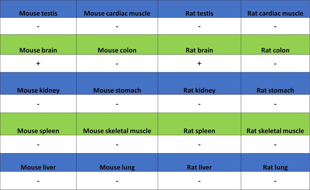
| Positive Sample | mouse brain, rat brain |
| Purification | Protein A |
| Concentration | 0.5 mg/ml |
| Conjugation | Unconjugated |
| Physical Appearance | Liquid |
| Storage Buffer | PBS, 40% Glycerol, 0.05% BSA, 0.02% sodium azide |
| Stability & Storage | 12 months from date of receipt / reconstitution, -20 °C as supplied |
Dilution
| application | dilution | species |
| WB | 1:1000 | Ms, Rt |
| IHC-P | 1:1000 | Ms, Rt |
| IF | 1:500 | Ms, Rt |
Background
Homer1 (Homer scaffold protein 1) is a postsynaptic density scaffold protein encoded by the HOMER1 gene, whose core function is to serve as a platform for neural signal transduction by recognizing and binding to various proline motif-containing signaling proteins, such as group I metabotropic glutamate receptors (mGluR1/5), inositol 1,4,5-trisphosphate receptors (IP3Rs), and ryanodine receptors (RyRs), through its N-terminal Ena/VASP homology 1 (EVH1) domain, thereby coupling cell surface receptors to intracellular calcium release channels and other effectors to regulate calcium homeostasis and synaptic plasticity. A major feature of this protein is the existence of multiple isoforms generated by alternative splicing, among which the long isoforms (such as Homer1b/c) contain a C-terminal coiled-coil domain that enables multimerization to stably scaffold protein complexes, whereas the short isoform Homer1a lacks this domain and, as an immediate early gene product, is rapidly induced upon neuronal activity and disrupts the complexes assembled by long isoforms in a dominant-negative manner, thereby initiating postsynaptic density remodeling—a mechanism considered critical for regulating long-term depression, synaptic homeostasis scaling during sleep, and stress susceptibility. Precisely because of its central role in excitatory synaptic transmission and plasticity, dysregulation of Homer1 function is closely associated with various neurological disorders, including epilepsy, drug addiction, and chronic inflammatory pain, and its gene polymorphisms are also linked to the risk of psychotic symptoms in Parkinson's disease; meanwhile, accumulating evidence indicates that Homer1 is aberrantly expressed in multiple cancers (such as hepatocellular carcinoma and colon cancer) and is associated with tumor prognosis and immune infiltration, highlighting its potential value as a diagnostic or prognostic biomarker.
Picture
Picture
Western Blot
WB result of Homer1 Recombinant Rabbit mAb
Primary antibody: Homer1 Recombinant Rabbit mAb at 1/1000 dilution
Lane 1: rat lung lysate 20 μg
Lane 2: rat brain lysate 20 μg
Negative control: mouse lung lysate
Secondary antibody: Goat Anti-Rabbit IgG (H+L), HRP conjugated at 1/10000 dilution
Predicted MW: 41 kDa
Observed MW: 46 kDa
This blot was developed with high sensitivity substrate
Immunohistochemistry
IHC shows positive staining in paraffin-embedded mouse cerebral cortex. Anti-Homer1 antibody was used at 1/1000 dilution, followed by a HRP Polymer for Rabbit IgG (ready to use). Counterstained with hematoxylin. Heat mediated antigen retrieval with Tris/EDTA buffer pH9.0 was performed before commencing with IHC staining protocol.
Negative control: IHC shows negative staining in paraffin-embedded mouse lung. Anti-Homer1 antibody was used at 1/1000 dilution, followed by a HRP Polymer for Rabbit IgG (ready to use). Counterstained with hematoxylin. Heat mediated antigen retrieval with Tris/EDTA buffer pH9.0 was performed before commencing with IHC staining protocol.
IHC shows positive staining in paraffin-embedded rat cerebral cortex. Anti-Homer1 antibody was used at 1/1000 dilution, followed by a HRP Polymer for Rabbit IgG (ready to use). Counterstained with hematoxylin. Heat mediated antigen retrieval with Tris/EDTA buffer pH9.0 was performed before commencing with IHC staining protocol.
Negative control: IHC shows negative staining in paraffin-embedded rat lung. Anti-Homer1 antibody was used at 1/1000 dilution, followed by a HRP Polymer for Rabbit IgG (ready to use). Counterstained with hematoxylin. Heat mediated antigen retrieval with Tris/EDTA buffer pH9.0 was performed before commencing with IHC staining protocol.
Immunofluorescence
IF shows positive staining in paraffin-embedded mouse cerebral cortex. Anti-Homer1 antibody was used at 1/500 dilution (Green) and incubated overnight at 4°C. Goat polyclonal Antibody to Rabbit IgG - H&L (Alexa Fluor® 488) was used as secondary antibody at 1/1000 dilution. Counterstained with DAPI (Blue). Heat mediated antigen retrieval with EDTA buffer pH9.0 was performed before commencing with IF staining protocol.
Negative control: IF shows negative staining in paraffin-embedded mouse lung. Anti-Homer1 antibody was used at 1/500 dilution and incubated overnight at 4°C. Goat polyclonal Antibody to Rabbit IgG - H&L (Alexa Fluor® 488) was used as secondary antibody at 1/1000 dilution. Counterstained with DAPI (Blue). Heat mediated antigen retrieval with EDTA buffer pH9.0 was performed before commencing with IF staining protocol.
IF shows positive staining in paraffin-embedded rat cerebral cortex. Anti-Homer1 antibody was used at 1/500 dilution (Green) and incubated overnight at 4°C. Goat polyclonal Antibody to Rabbit IgG - H&L (Alexa Fluor® 488) was used as secondary antibody at 1/1000 dilution. Counterstained with DAPI (Blue). Heat mediated antigen retrieval with EDTA buffer pH9.0 was performed before commencing with IF staining protocol.
Negative control: IF shows negative staining in paraffin-embedded rat lung. Anti-Homer1 antibody was used at 1/500 dilution and incubated overnight at 4°C. Goat polyclonal Antibody to Rabbit IgG - H&L (Alexa Fluor® 488) was used as secondary antibody at 1/1000 dilution. Counterstained with DAPI (Blue). Heat mediated antigen retrieval with EDTA buffer pH9.0 was performed before commencing with IF staining protocol.