For reconstitution, pre-warm the kit to room temperature; add 100 µL of DMSO to one vial of Fixable Viability Dye 620 and mix until fully dissolved. After reconstitution with DMSO, store the stock solution at -20°C in small aliquots. Use up within 3 months. Repeated freezing and thawing is not recommended.
For recommended usage, add 1 µl of Fixable Viability Dye 620 Stock Solution for each 2~5 ml of cell suspension/PBS (1:2000~1:5000) and vortex immediately. It is recommended that the reagent be titrated for optimal performance for each application.
Please refer to the protocol for details.
Fixable Viability Dye 620
Fixable Viability Dye 620
Price:
Regular price
$105 USD
Regular price
Sale price
$105 USD
Unit price
per
For shipping services or bulk orders, you may request a quotation.
Secure checkout with

View full details
Product Details
Product Details
Product Specification
| Stability & Storage | 12 months from date of receipt, -20 °C as supplied |
Background
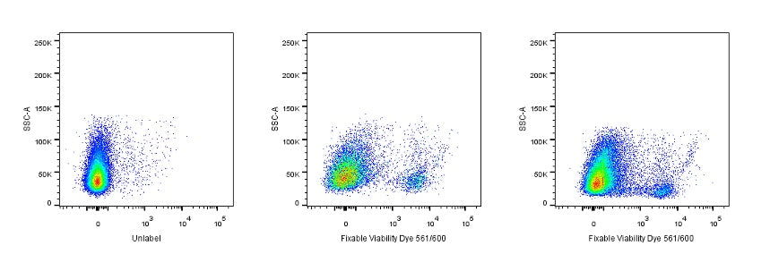
Fixable Viability Dye 620 is excited by the Yellow laser (with an excitation maximum of 561 nm) and has fluorescence emission maximum at 600 nm, Fixable Viability Dye 620 has similar emission to PE-CF594, PI. Fixable Viability Dye 620 can intracellular diffusion of the dye and covalent binding to higher overall concentrations of amines, Each lot of this product is quality control tested, is useful for discrimination of viable from non-viable mammalian cells in multicolor flow cytometric applications. Each lot of this product is quality control tested. The labeled cells can be fixed with formaldehyde for downstream decontamination, freezing and/or permeabilization and subsequent intracellular staining while maintaining stable viability stain fluorescence.
Picture
Picture
Contents & Protocol
FC
Rabbit splenocytes were stained with Fixable Viability Dye 620 and analyzed before fixation (Middle Panels) or after fixation (Right Panels). Cells alone, without Fixable Viability Dye 620 staining (Left Panels). Flow cytometry and data analysis were performed using BD FACSymphony™ A1 and FlowJo™ software.