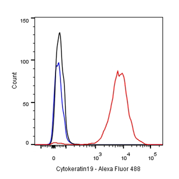
Flow cytometric analysis of MCF7 cells labelling Cytokeratin 19 antibody at 1/500 dilution/ (red) compared with a Rabbit monoclonal IgG (Black) isotype control and an unlabelled control (cells without incubation with primary antibody and secondary antibody) (Blue). Goat Anti-Rabbit IgG Alexa Fluor® 488 at 1/1000 dilution was used as the secondary antibody.
Cytokeratin 19 Recombinant Rabbit mAb,PBS Only (SDT-107-19)
Cytokeratin 19 Recombinant Rabbit mAb,PBS Only (SDT-107-19)
Price:
Regular price
$0 USD
Regular price
Sale price
$0 USD
Unit price
per
For shipping services or bulk orders, you may request a quotation.
Secure checkout with

View full details
Product Details
Product Details
Product Specification
| Host | Rabbit |
| Antigen | Cytokeratin 19 |
| Synonyms | KRT19, CK-19 |
| Immunogen | Synthetic Peptide |
| Location | Cell membrane |
| Accession | P08727 |
| Clone Number | SDT-107-19 |
| Antibody Type | Rabbit mAb |
| Application | WB, IHC-P, ICC, FCM |
| Reactivity | Hu, Ms, Rt |
| Purification | Protein A |
| Concentration | 1 mg/ml |
| Physical Appearance | Liquid |
| Storage Buffer | PBS |
| Stability & Storage | 12 months from date of receipt, 4°C as supplied |
Dilution
| application | dilution | species |
| WB | 1:500 | |
| IHC-P | 1:1000 | |
| FCM | 1:500 | |
| ICC | 1:250 |
Background
Cytokeratin 19 is a member of the keratin family. The keratins are intermediate filament proteins responsible for the structural integrity of epithelial cells and are subdivided into cytokeratins and hair keratins. Keratin 19 is a type I keratin. The type I cytokeratins consist of acidic proteins which are arranged in pairs of heterotypic keratin chains. Unlike its related family members, this smallest known acidic cytokeratin is not paired with a basic cytokeratin in epithelial cells. It is specifically found in the periderm, the transiently superficial layer that envelops the developing epidermis. KRT19 is also known as Cyfra 21-1.Due to its high sensitivity, KRT19 is the most used marker for the RT-PCR-mediated detection of tumor cells disseminated in lymph nodes, peripheral blood, and bone marrow of breast cancer patients.
Picture
Picture
Validation Data
Western Blot
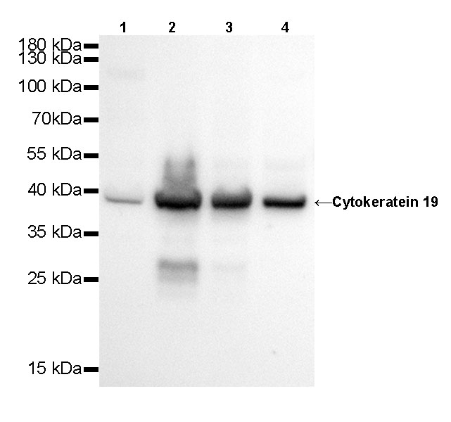
WB result of Cytokeratin 19 Rabbit mAb
Primary antibody: Cytokeratin 19 Rabbit mAb at 1/500 dilution
Lane 1: Hela whole cell lysate 20 µg
Lane 2: MCF7 whole cell lysate 20 µg
Lane 3: HT-29 whole cell lysate 20 µg
Lane 4: HepG2 whole cell lysate 20 µg
Secondary antibody: Goat Anti-Rabbit IgG, (H+L), HRP conjugated at 1/10000 dilution
Predicted MW: 41 kDa
Observed MW: 39 kDa
Exposure time: 20s
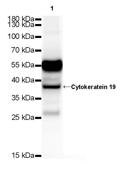
WB result of Cytokeratin 19 Rabbit mAb
Primary antibody: Cytokeratin 19 Rabbit mAb at 1/500 dilution
Lane 1: mouse kidney lysate 20 µg
Secondary antibody: Goat Anti-Rabbit IgG, (H+L), HRP conjugated at 1/10000 dilution
Predicted MW: 41 kDa
Observed MW: 39 kDa
Exposure time: 120 s
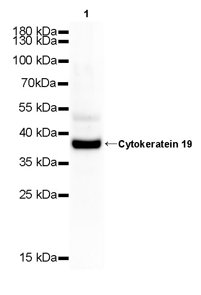
WB result of Cytokeratin 19 Rabbit mAb
Primary antibody: Cytokeratin 19 Rabbit mAb at 1/500 dilution
Lane 1: rat kidney whole cell lysate 20 µg
Secondary antibody: Goat Anti-Rabbit IgG, (H+L), HRP conjugated at 1/10000 dilution
Predicted MW: 41 kDa
Observed MW: 39 kDa
Exposure time: 120 s
Immunohistochemistry
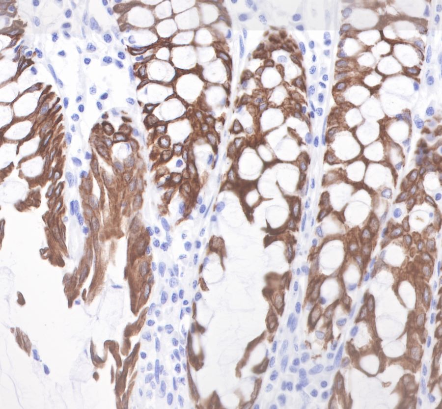
IHC shows positive staining in paraffin-embedded human colon.
Anti-Cytokeratin19 antibody was used at 1/1000 dilution, followed by a Goat Anti-Rabbit IgG H&L (HRP) ready to use.
Counterstained with hematoxylin.
Heat mediated antigen retrieval with Tris/EDTA buffer pH9.0 was performed before commencing with IHC staining protocol.
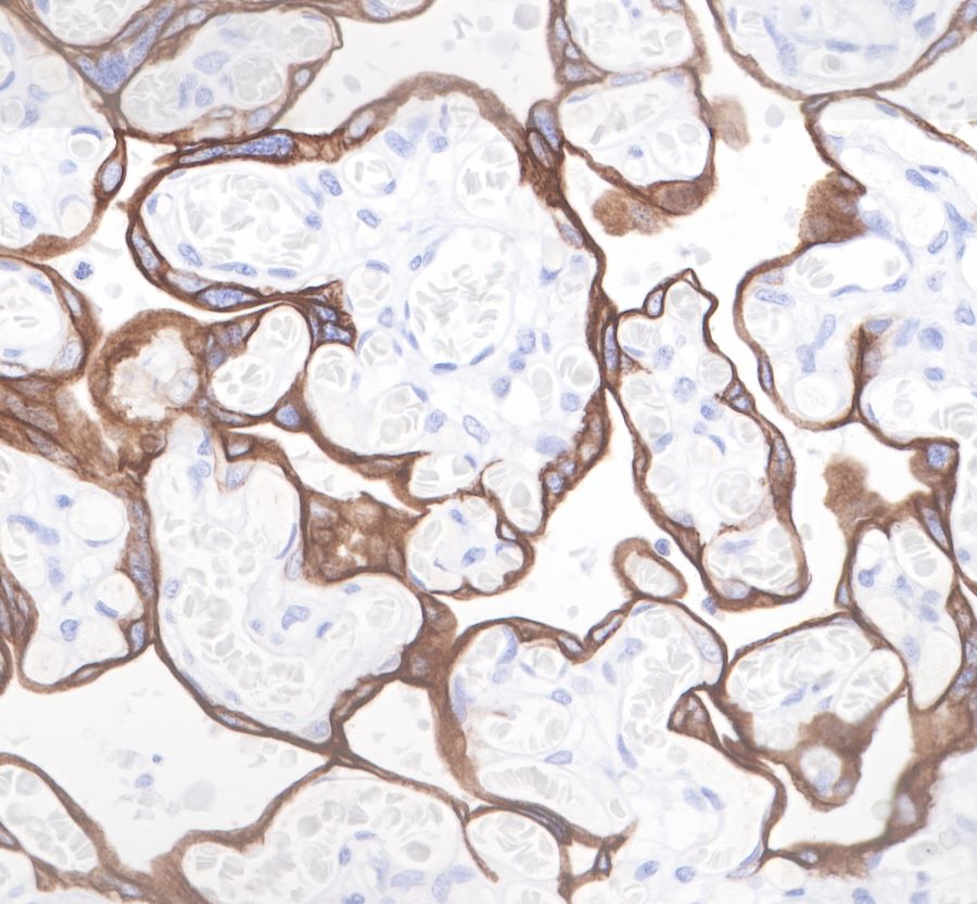
IHC shows positive staining in paraffin-embedded human placenta.
Anti-Cytokeratin19 antibody was used at 1/1000 dilution, followed by a Goat Anti-Rabbit IgG H&L (HRP) ready to use.
Counterstained with hematoxylin.
Heat mediated antigen retrieval with Tris/EDTA buffer pH9.0 was performed before commencing with IHC staining protocol.
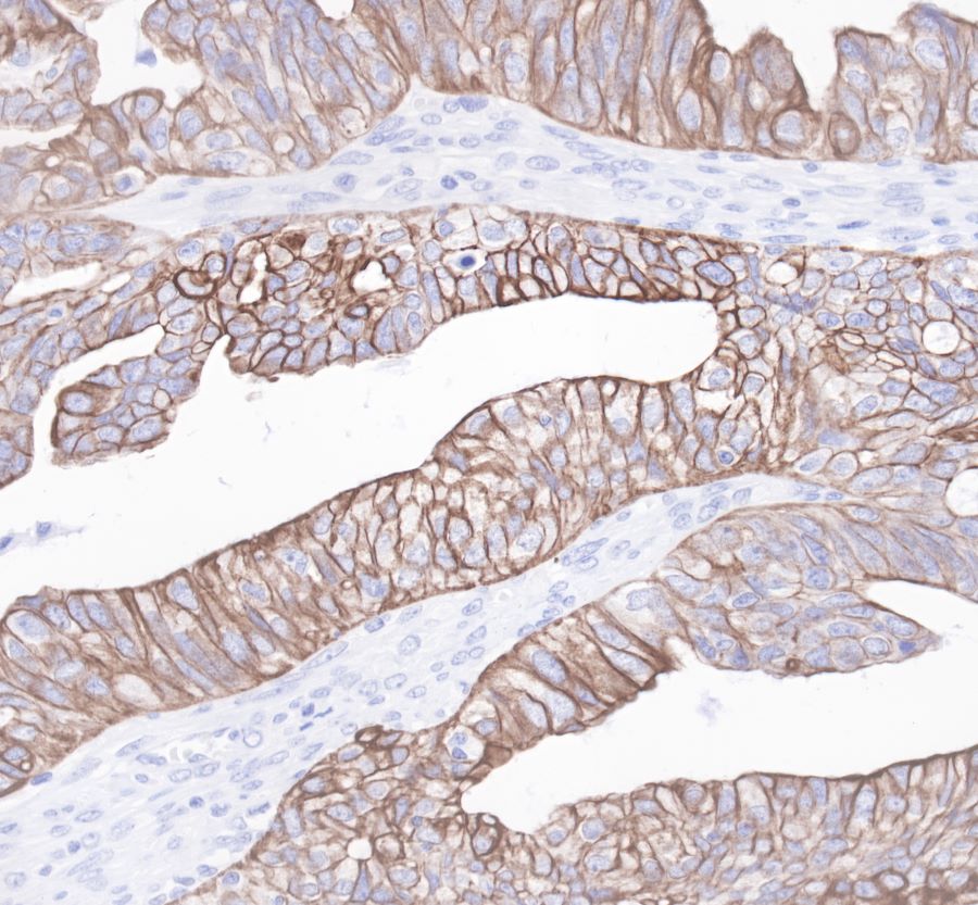
IHC shows positive staining in paraffin-embedded human ovarian cancer. Anti-Cytokeratin19 antibody was used at 1/1000 dilution, followed by a Goat Anti-Rabbit IgG H&L (HRP) ready to use.
Counterstained with hematoxylin.
Heat mediated antigen retrieval with Tris/EDTA buffer pH9.0 was performed before commencing with IHC staining protocol.
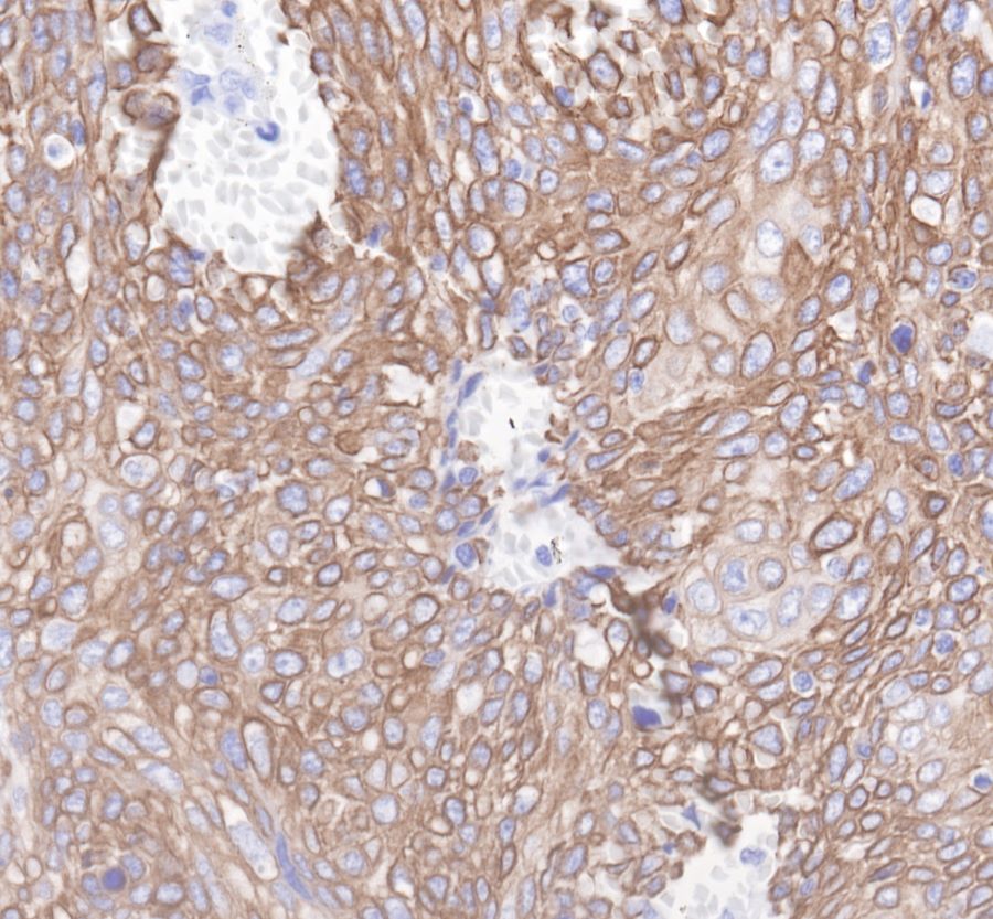
IHC shows positive staining in paraffin-embedded human cervix cancer. Anti-Cytokeratin19 antibody was used at 1/1000 dilution, followed by a Goat Anti-Rabbit IgG H&L (HRP) ready to use.
Counterstained with hematoxylin.
Heat mediated antigen retrieval with Tris/EDTA buffer pH9.0 was performed before commencing with IHC staining protocol.
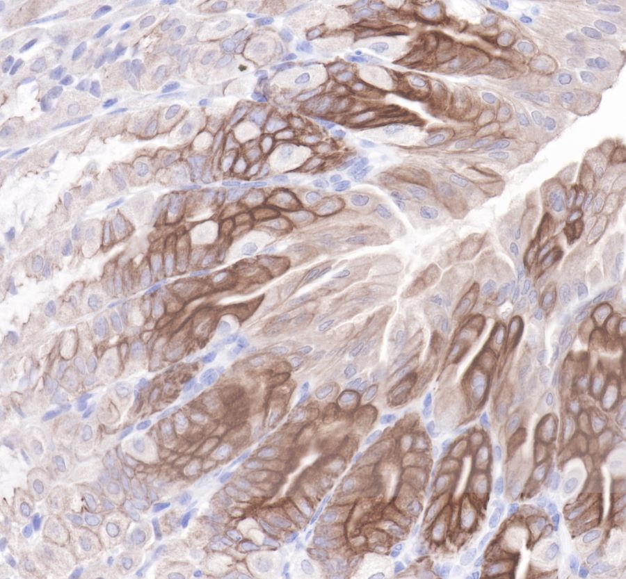
IHC shows positive staining in paraffin-embedded mouse stomach.
Anti-Cytokeratin19 antibody was used at 1/1000 dilution, followed by a Goat Anti-Rabbit IgG H&L (HRP) ready to use.
Counterstained with hematoxylin.
Heat mediated antigen retrieval with Tris/EDTA buffer pH9.0 was performed before commencing with IHC staining protocol.
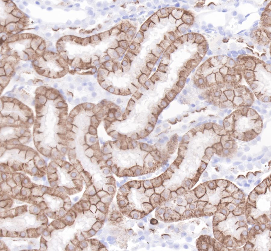
IHC shows positive staining in paraffin-embedded rat kidney.
Anti-Cytokeratin19 antibody was used at 1/1000 dilution, followed by a Goat Anti-Rabbit IgG H&L (HRP) ready to use.
Counterstained with hematoxylin.
Heat mediated antigen retrieval with Tris/EDTA buffer pH9.0 was performed before commencing with IHC staining protocol.
Immunocytochemistry
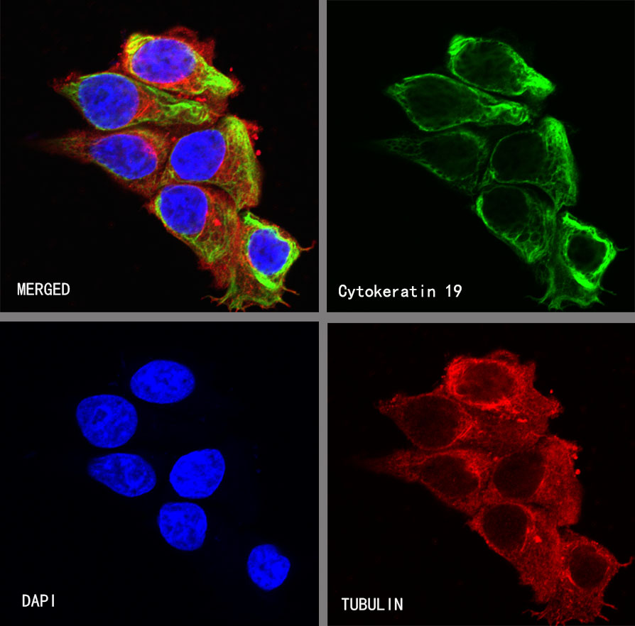
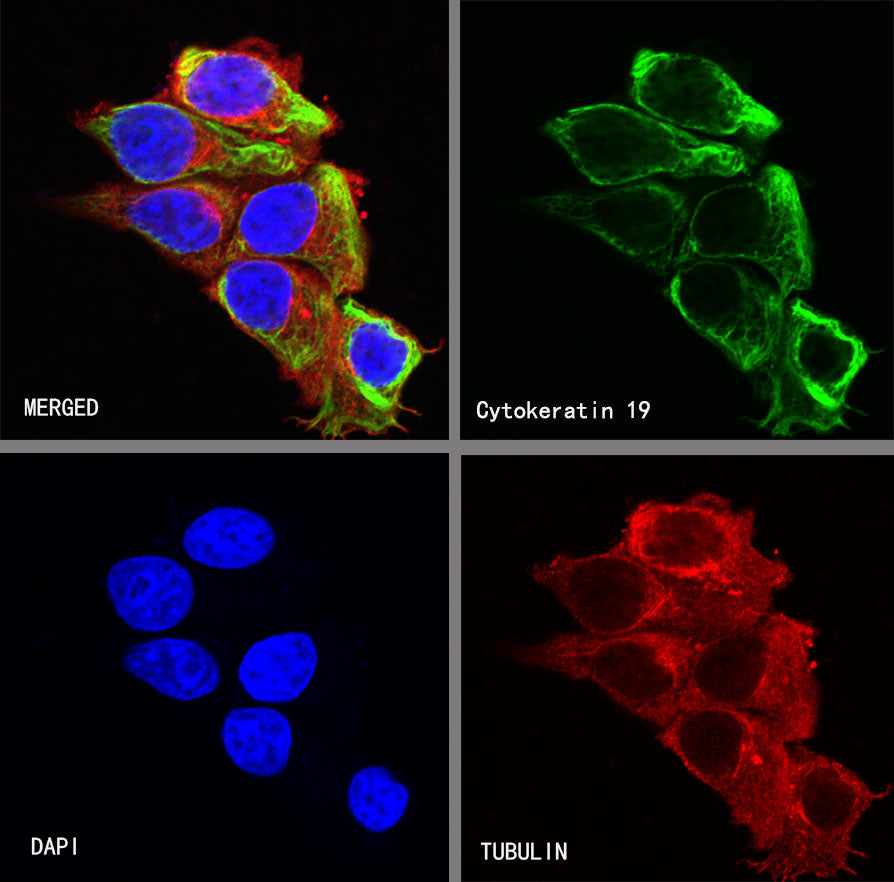
ICC shows positive staining in MCF7 cells. Anti-Cytokeratin 19 antibody was used at 1/250 dilution (Green) and incubated overnight at 4°C. Goat polyclonal Antibody to Rabbit IgG - H&L (Alexa Fluor® 488) was used as secondary antibody at 1/1000 dilution. The cells were fixed with 4% PFA and permeabilized with 0.1% PBS-Triton X-100. Nuclei were counterstained with DAPI (Blue). Counterstain with tubulin (red).