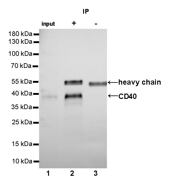
Flow cytometric analysis of Jurkat (left) / Ramos (right) cells labelling CD40 antibody at 1/500 dilution (0.1ug)/ (red) compared with a Rabbit monoclonal IgG (Black) isotype control and an unlabelled control (cells without incubation with primary antibody and secondary antibody) (Blue). Goat Anti-Rabbit IgG Alexa Fluor® 488 was used as the secondary antibody. Negative control: Jurkat
CD40 Recombinant Rabbit mAb,PBS Only (SDT-145-45)
CD40 Recombinant Rabbit mAb,PBS Only (SDT-145-45)
Price:
Regular price
$0 USD
Regular price
Sale price
$0 USD
Unit price
per
For shipping services or bulk orders, you may request a quotation.
Secure checkout with

View full details
Product Details
Product Details
Product Specification
| Host | Rabbit |
| Antigen | CD40 |
| Synonyms | B-cell surface antigen CD40,Bp50,CD40L receptor,CDw40 |
| Immunogen | Recombinant Protein |
| Location | Secreted, Cell membrane |
| Accession | P25942 |
| Clone Number | SDT-145-45 |
| Antibody Type | Rabbit mAb |
| Application | WB, IHC-P, FCM, IP |
| Reactivity | Hu |
| Purification | Protein A |
| Concentration | 1 mg/ml |
| Physical Appearance | Liquid |
| Storage Buffer | PBS |
| Stability & Storage | 12 months from date of receipt, 4°C as supplied |
Dilution
| application | dilution | species |
| WB | 1:1000 | |
| IHC-P | 1:100-500 | |
| FCM | 1:500 | |
| IP | 1:25 |
Background
The CD40 is a type I transgender protein that is transmitted in the immune system's B cells and special antigen to the surface of the cells and many non -hematopoietic cell types and cancer cells. It belongs to the TNF receptor super family.CD40 is expressed in B cells, foam dendritic cells, dendritic cells, activated monocytes, macrophages, endothelial cells, vascular smooth muscle cells, and several tumor cell lines. CD40 interacts with CD40 Ligand (CD40L/TNFSF5).CD40/CD40L is essential for the beginning and progress of cells and body fluid adaptive immunity, and it is an important area of concern in the study of tumor immunology, neurodegeneration, vascular disease and inflammatory diseases.
Picture
Picture
Validation Data
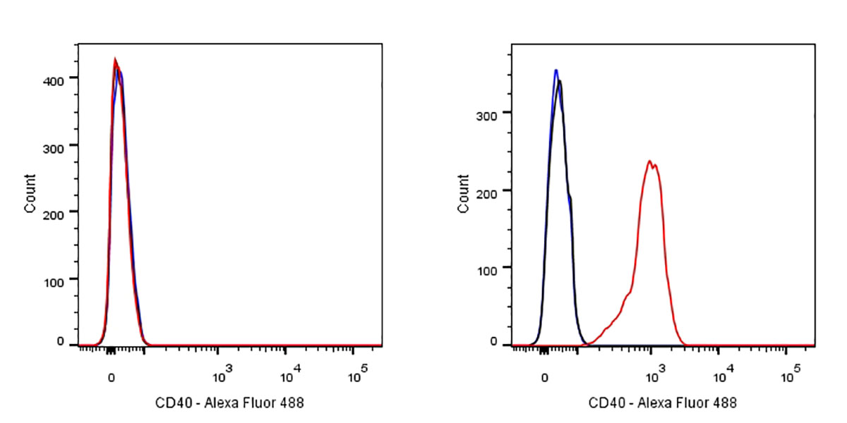
Western Blot

WB result of CD40 Rabbit mAb
Primary antibody:CD40 Rabbit mAb at 1/1000 dilution
Lane 1: Jurkat whole cell lysate 20 µg
Lane 2: Raji whole cell lysate 20 µg
Lane 3: Ramos whole cell lysate 20 µg
Lane 4: Daudi whole cell lysate 20 µg
Negative control: Jurkat whole cell lysate
Secondary antibody: Goat Anti-Rabbit IgG, (H+L), HRP conjugated at 1/10000 dilution
Predicted MW: 42 kDa
Observed MW: 39 kDa
Exposure time: 40s
IP
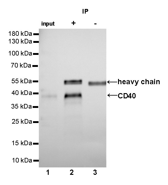
CD40 Rabbit mAb at 1/25 dilution (2µg) immunoprecipitating CD40 in 0.4mg Raji whole cell lysate.
Western blot was performed on the immunoprecipitate using CD40 Rabbit mAb at 1/1000 dilution.
Secondary antibody (HRP) for IP was used at 1/400 dilution.
Lane 1: Raji whole cell lysate 10µg (input)
Lane 2 (+): CD40 Rabbit mAb IP in Raji whole cell lysate
Lane 3 (-): Rabbit monoclonal IgG IP in Raji whole cell lysate
Predicted MW: 42 kDa
Observed MW: 39 kDa
Exposure time: 50s
Immunohistochemistry
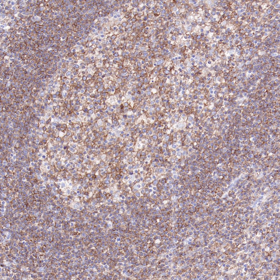
IHC shows positive staining in paraffin-embedded human tonsil.
Anti-CD40 antibody was used at 1/100 dilution, followed by a Goat Anti-Rabbit IgG H&L (HRP) ready to use. Counterstained with hematoxylin.
Heat mediated antigen retrieval with Tris/EDTA buffer pH9.0 was performed before commencing with IHC staining protocol.

IHC shows positive staining in paraffin-embedded human spleen.
Anti-CD40 antibody was used at 1/100 dilution, followed by a Goat Anti-Rabbit IgG H&L (HRP) ready to use. Counterstained with hematoxylin.
Heat mediated antigen retrieval with Tris/EDTA buffer pH9.0 was performed before commencing with IHC staining protocol.
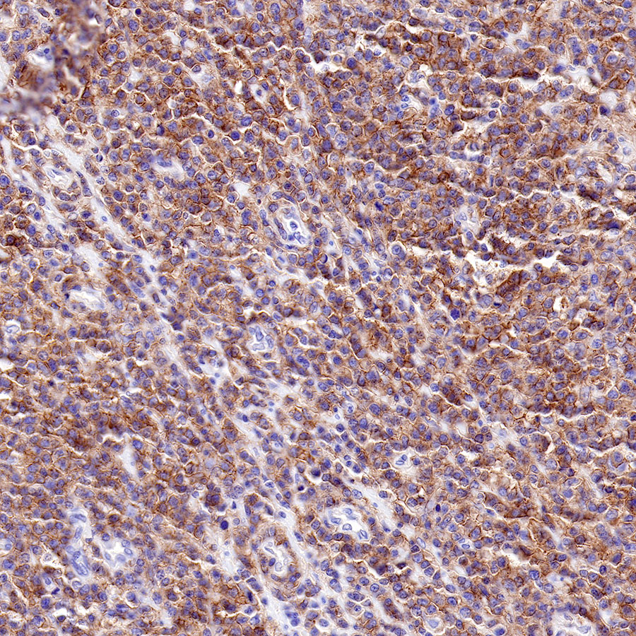
IHC shows positive staining in paraffin-embedded human diffuse large B-cell lymphoma.
Anti-CD40 antibody was used at 1/500 dilution, followed by a Goat Anti-Rabbit IgG H&L (HRP) ready to use. Counterstained with hematoxylin.
Heat mediated antigen retrieval with Tris/EDTA buffer pH9.0 was performed before commencing with IHC staining protocol.
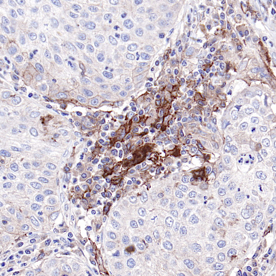
IHC shows positive staining in paraffin-embedded human lung squamous cancer.
Anti-CD40 antibody was used at 1/100 dilution, followed by a Goat Anti-Rabbit IgG H&L (HRP) ready to use. Counterstained with hematoxylin.
Heat mediated antigen retrieval with Tris/EDTA buffer pH9.0 was performed before commencing with IHC staining protocol.