CellXViva Mouse T cell Activation Kit
CellXViva Mouse T cell Activation Kit

Product Details
Product Details
Product Specification
| Species | Mouse |
| Reconstitution | IL-2 Protein, Mouse: Reconstitute at 0.1-1 mg/ml according to the size in ultrapure water after rapid centrifugation. |
| Stability & Storage | -20 to -80℃. Please avoid repeated freeze-thaw cycles. |
Components
Contain |
Formulation |
Reference dosage |
S Size (200mL system) |
M Size (1L system) |
L Size (2L system) |
IL-2 Protein, Mouse |
Lyophilized Powder |
10 ng/mL |
5ug |
10ug |
20ug |
NA/LE Rat anti-mouse CD3 Recombinant mAb |
Liquid |
10 µg/mL |
2mg |
10mg |
20mg |
NA/LE Syrian Hamster anti-mouse CD28 mAb |
Liquid |
2 µg/mL |
0.5mg |
2mg |
4mg |
Protocol
Complete medium formulation for reference:
Component |
Final Concentration |
RPMI-1640 |
90% |
Fetal Bovine Serum |
10% |
Sodium Pyruvate100x |
1x |
L-Glutamine100x |
1x |
HEPES |
1x |
Penicillin-Streptomycin |
1x |
β-Mercaptoethanol |
55 μM |
Protocol 1: Anti-Mouse CD3 Monoclonal Antibody Coating Method
1. Dilute NA/LE Rat anti-mouse CD3 Recombinant mAb into PBS to a final concentration of 10 µg/mL.
2. Add the diluted NA/LE Rat anti-mouse CD3 Recombinant mAb to a 24-well plate at 1 mL per well for coating.
2. Incubate for 2 hours at 37°C with 5% CO₂, or overnight at 4°C.
4. Remove the NA/LE Rat anti-mouse CD3 Recombinant mAb from the 24-well plate.
5. Prepare a single-cell suspension from mouse spleen. For T cell activation, cells should be resuspended in complete medium at a concentration of (1-3)×10⁶ cells/mL. For a 24-well plate, a concentration of 1×10⁶ cells/mL is recommended.
6. Prepare fully supplemented T cell medium in a V-bottom tube, and add the following cytokines and antibodies to the cell suspension from step 6:
- 2µg/mL NA/LE Syrian Hamster anti-mouse CD28 mAb
- 10ng/mL IL-2 Protein, Mouse
7. Transfer the required amount of cells to an appropriate cell culture plate to a final density of (1-1.5)×10⁶ cells/mL/cm².
8. Incubate for 2 days at 37°C with 5% CO₂.
9. After 2 days of culture, check cell health and record images.
10. Centrifuge the cell suspension at 400 × g for 5 minutes, discard the supernatant. Wash the cell pellet with PBS.
11. Centrifuge the cell suspension at 400 × g for 5 minutes, discard the supernatant. Resuspend the cells in 0.5 mL of 1% Bovine Serum Albumin (BSA) and count the cells. Assess cell viability, requiring >95% viability.
12. Block 1×10⁶ cells with 10 µg of mouse IgG antibody. Incubate on ice for 30 minutes.
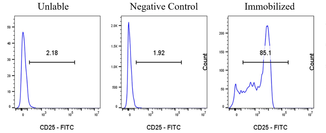
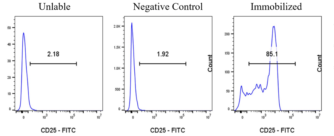
13. Add FITC Rat anti-Mouse CD25 Antibody (S-R441) (S0B5215) according to the instructions, and incubate at 4°C for 30 minutes.
14. Centrifuge the cell suspension at 400 × g for 5 minutes, and wash the cell pellet twice with PBS containing 1% BSA.
15. Resuspend the cell pellet in 200 µL PBS for flow cytometry analysis (requires >10,000 cells).
Picture
Picture
FC