WB result of SMAD2/3 Recombinant Rabbit mAb
Primary antibody: SMAD2/3 Recombinant Rabbit mAb at 1/1000 dilution
Lane 1: HeLa whole cell lysate 20 µg
Lane 2: HCT 116 whole cell lysate 20 µg
Secondary antibody: Goat Anti-rabbit IgG, (H+L), HRP conjugated at 1/10000 dilution
Predicted MW: 48, 52 kDa
Observed MW: 52, 60 kDa
SMAD2/3 Recombinant Rabbit mAb (S-5467)
SMAD2/3 Recombinant Rabbit mAb (S-5467)
Price:
Regular price
$100 USD
Regular price
Sale price
$100 USD
Unit price
per
For shipping services or bulk orders, you may request a quotation.
Secure checkout with

View full details
Product Details
Product Details
Product Specification
| Host | Rabbit |
| Antigen | SMAD2/3 |
| Location | Cytoplasm, Nucleus |
| Accession | Q15796、P84022 |
| Clone Number | S-5467 |
| Antibody Type | Recombinant mAb |
| Isotype | IgG |
| Application | WB, ChIP |
| Reactivity | Hu, Ms |
| Positive Sample | HeLa, HCT 116, C2C12 |
| Predicted Reactivity | Rt, Mk |
| Purification | Protein A |
| Concentration | 0.5 mg/ml |
| Conjugation | Unconjugated |
| Physical Appearance | Liquid |
| Storage Buffer | PBS, 40% Glycerol, 0.05% BSA, 0.02% sodium azide |
| Stability & Storage | 12 months from date of receipt / reconstitution, -20 °C as supplied |
Dilution
| application | dilution | species |
| WB | 1:1000-1:2000 | Hu, Ms |
| ChIP | 1:20-1:50 | Hu |
Background
SMAD2 and SMAD3 are two highly homologous and functionally closely related mediator signal transduction proteins in the TGF-β signaling pathway. Structurally, they both consist of an MH1 (MAD homology 1) domain, an MH2 domain, and a linker region in between, with the MH1 domain responsible for sequence-specific DNA binding and nuclear localization, while the MH2 domain mediates interactions with various proteins including receptors, SMAD4, and transcriptional cofactors. In the canonical signaling pathway, upon TGF-β ligand binding to its receptor, the receptor-regulated SMADs (R-SMADs), namely SMAD2 and SMAD3, are phosphorylated and activated, forming heterotrimeric complexes with the common SMAD4 and translocating into the nucleus. Once in the nucleus, these complexes bind to SMAD-binding elements on DNA and, in coordination with numerous transcriptional coactivators or corepressors, initiate or repress the expression of specific target genes, thereby regulating a broad spectrum of biological processes including cell proliferation, differentiation, apoptosis, migration, and extracellular matrix remodeling. Although SMAD2 and SMAD3 belong to the same R-SMAD family, they exhibit significant functional differences: SMAD3 can bind DNA directly, whereas SMAD2 requires cooperation with SMAD4 or other cofactors to stably associate with DNA; moreover, SMAD3 plays a more prominent role in inducing apoptosis and inhibiting cell growth, while SMAD2 is more focused on regulating embryonic stem cell fate. Dysregulation of these two signaling pathways is associated with various diseases, where their functional abnormalities can promote tissue fibrosis and malignant tumor progression through overactivation, or lead to developmental defects and immune dysregulation through loss of function.
Picture
Picture
Western Blot
WB result of SMAD2/3 Recombinant Rabbit mAb
Primary antibody: SMAD2/3 Recombinant Rabbit mAb at 1/1000 dilution
Lane 1: C2C12 whole cell lysate 20 µg
Secondary antibody: Goat Anti-rabbit IgG, (H+L), HRP conjugated at 1/10000 dilution
Predicted MW: 48, 52 kDa
Observed MW: 52, 60 kDa
ChIP
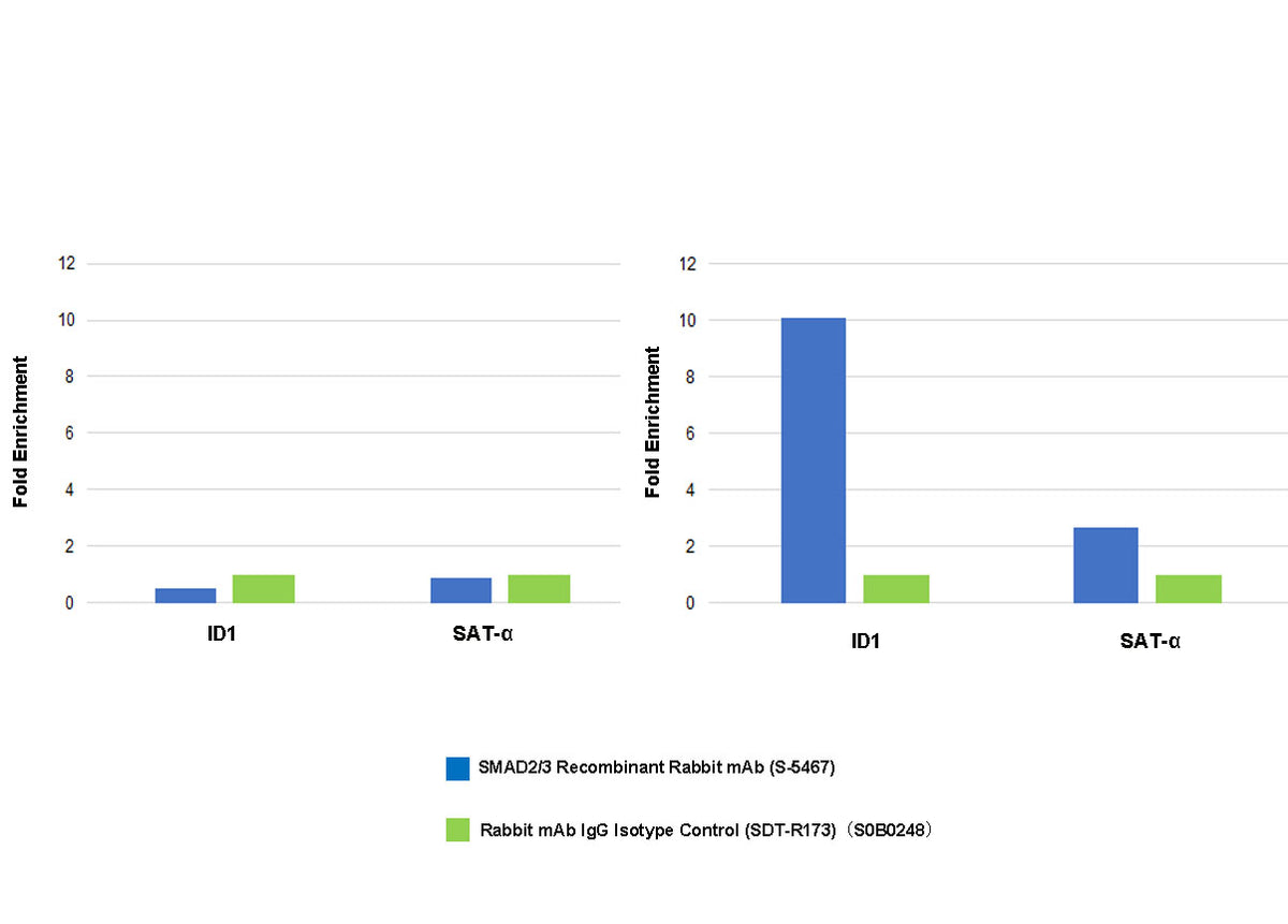
Chromatin immunoprecipitation (ChIP) was performed on HaCaT+Htgf-beta3(7ng/ml, 1h)(-)(left), HaCaT+Htgf-beta3(7ng/ml, 1h)(+)(right) cross - linked with 1% formaldehyde for 10 min, then chromatin was fragmented by sonication.
Parallel reactions used SMAD2/3 Recombinant Rabbit mAb (S-5467) and Rabbit mAb IgG Isotype Control (SDT-R173) at 1:50 for immunoprecipitation.
Post - immunoprecipitation, both samples were washed, eluted, and cross - links reversed. Purified DNA was analyzed by qPCR.
qPCR showed the enrichment of ID1 and SAT-α in SMAD2/3 Recombinant Rabbit mAb (S-5467)-immunoprecipitated sample.