WB result of PTGES Recombinant Rabbit mAb
Primary antibody: PTGES Recombinant Rabbit mAb at 1/1000 dilution
Lane 1: A549 whole cell lysate 20 µg
Lane 2: HeLa whole cell lysate 20 µg
Secondary antibody: Goat Anti- rabbit IgG, (H+L), HRP conjugated at 1/10000 dilution
Predicted MW: 17 kDa
Observed MW: 17 kDa
PTGES Recombinant Rabbit mAb (S-4351-18)
PTGES Recombinant Rabbit mAb (S-4351-18)
Price:
Regular price
$100 USD
Regular price
Sale price
$100 USD
Unit price
per
For shipping services or bulk orders, you may request a quotation.
Secure checkout with

View full details
Product Details
Product Details
Product Specification
| Host | Rabbit |
| Antigen | PTGES |
| Synonyms | Prostaglandin E synthase; Glutathione peroxidase PTGES; Glutathione transferase PTGES; Microsomal glutathione S-transferase 1-like 1 (MGST1-L1); Microsomal prostaglandin E synthase 1 (MPGES-1); p53-induced gene 12 protein; MGST1L1; MPGES1; PGES; PIG12 |
| Immunogen | Synthetic Peptide |
| Location | Cytoplasm |
| Accession | O14684 |
| Clone Number | S-4351-18 |
| Antibody Type | Recombinant mAb |
| Isotype | IgG |
| Application | WB, IHC-P |
| Reactivity | Hu, Ms, Rt |
| Purification | Protein A |
| Concentration | 0.5 mg/ml |
| Conjugation | Unconjugated |
| Physical Appearance | Liquid |
| Storage Buffer | PBS, 40% Glycerol, 0.05% BSA, 0.02% sodium azide |
| Stability & Storage | 12 months from date of receipt / reconstitution, -20 °C as supplied |
Dilution
| application | dilution | species |
| WB | 1:1000 | Hu, Ms, Rt |
| IHC-P | 1:1000 | Hu |
Background
PTGES, also known as prostaglandin E synthase (or microsomal prostaglandin E synthase 1, mPGES-1), is a key enzyme composed of 152 amino acids encoded by the PTGES gene in humans, and belongs to the MAPEG family of membrane-associated proteins. As the terminal rate-limiting enzyme in the arachidonic acid metabolic pathway, PTGES is an integral membrane protein primarily localized to the endoplasmic reticulum and nuclear membranes, functioning as a homotrimer. Its core function is to isomerize prostaglandin H₂ (PGH₂), the product of cyclooxygenases (COX-1/2), into prostaglandin E₂ (PGE₂)—a lipid mediator that plays a central role in inflammation, fever, and pain—in a glutathione (GSH)-dependent manner. The expression of PTGES is inducible and can be significantly upregulated by pro-inflammatory cytokines such as interleukin-1β (IL1B) as well as by the tumor suppressor protein p53 (TP53). In addition to its primary PGE₂ synthase activity, PTGES also exhibits certain glutathione transferase and peroxidase activities. Given its critical role in inflammatory responses, PTGES is widely regarded as a potential drug target for the treatment of rheumatoid arthritis, pain, and various cancers, and the development of specific inhibitors targeting this enzyme is a major focus of anti-inflammatory drug research.
Picture
Picture
Western Blot
Immunohistochemistry
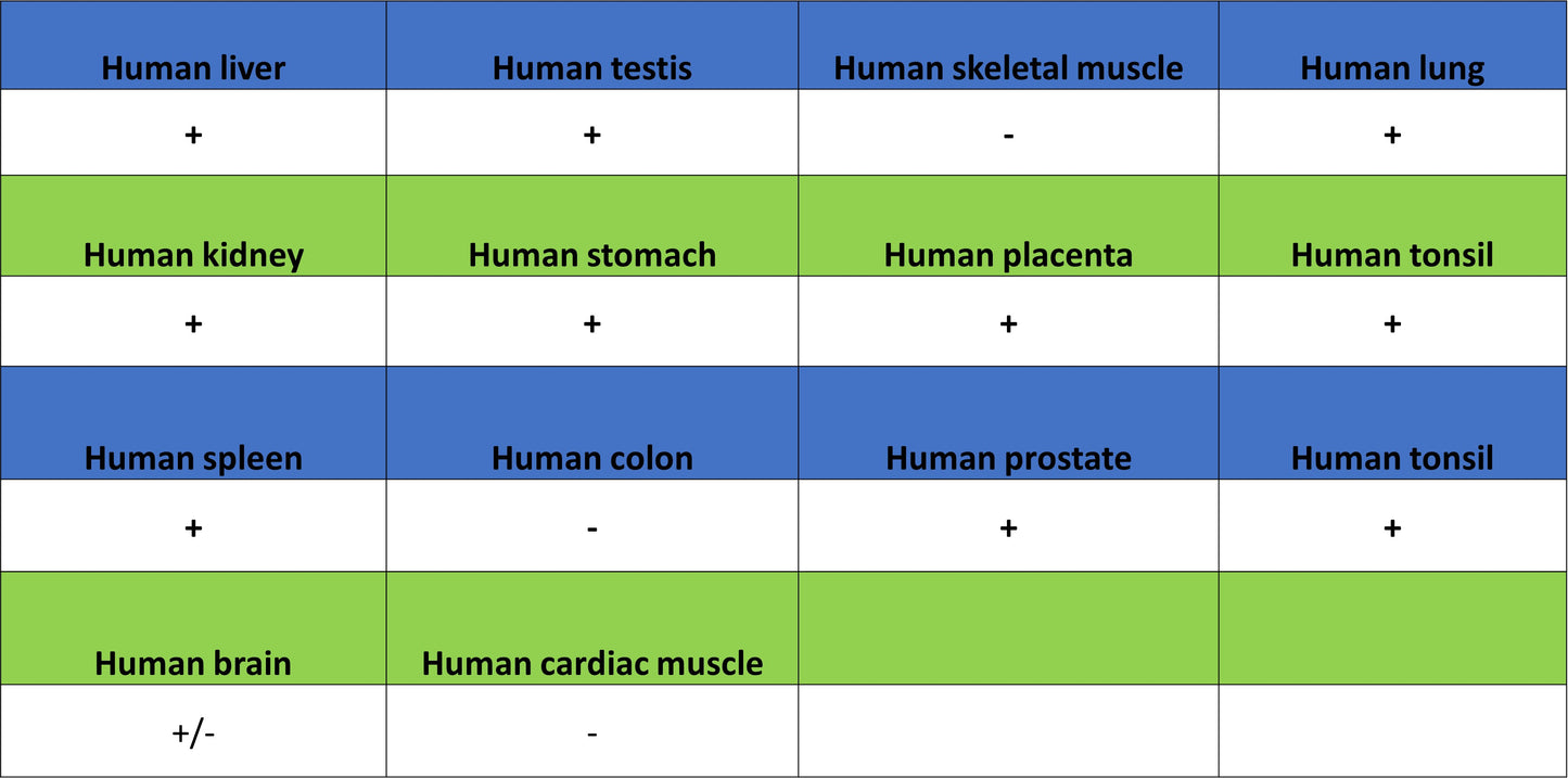
IHC shows positive staining in paraffin-embedded human placenta. Anti-PTGES antibody was used at 1/1000 dilution, followed by a HRP Polymer for Rabbit IgG (ready to use). Counterstained with hematoxylin. Heat mediated antigen retrieval with Tris/EDTA buffer pH9.0 was performed before commencing with IHC staining protocol.
IHC shows positive staining in paraffin-embedded human testis. Anti-PTGES antibody was used at 1/1000 dilution, followed by a HRP Polymer for Rabbit IgG (ready to use). Counterstained with hematoxylin. Heat mediated antigen retrieval with Tris/EDTA buffer pH9.0 was performed before commencing with IHC staining protocol.
IHC shows positive staining in paraffin-embedded human tonsil. Anti-PTGES antibody was used at 1/1000 dilution, followed by a HRP Polymer for Rabbit IgG (ready to use). Counterstained with hematoxylin. Heat mediated antigen retrieval with Tris/EDTA buffer pH9.0 was performed before commencing with IHC staining protocol.
IHC shows positive staining in paraffin-embedded human colon cancer. Anti-PTGES antibody was used at 1/1000 dilution, followed by a HRP Polymer for Rabbit IgG (ready to use). Counterstained with hematoxylin. Heat mediated antigen retrieval with Tris/EDTA buffer pH9.0 was performed before commencing with IHC staining protocol.