
Kanamycin Residue Detection Kit
Kanamycin Residue Detection Kit
Price:
Regular price
$697 USD
Regular price
Sale price
$697 USD
Unit price
per
For shipping services or bulk orders, you may request a quotation.
Secure checkout with

View full details
Product Details
Product Details
Product Specification
| Usage |
I. Preparation: 1 Kit preparation Each component of the kit was allowed to equilibrate at room temperature 30min After starting the operation. 2 Consumables and equipment that need to be provided by yourself ( 1 ) microplate reader, thermostatic shaker (or thermostatic incubator), plate washer, vortex shaker, timer; ( 2 ) High-precision pipettes and disposable tips ( 0.5-10uL 、 10-100uL 、 30-300uL 、 100-1000uL ); ( 3 ) deionized water (used to prepare the lotion ( 1× )); ( 4 ) absorbent paper, EP tubes, disposable gloves; 3 Reagent preparation ( 1 ) Lotion ( 1× ) Formulation Take the lotion ( 20× ) 1 Portions, plus 19 Part of deionized water to prepare a working concentration washing solution ( 1× )。 Lotion ( 20× If there is crystal formation, it should be placed at room temperature or 37℃ Shake gently in the water bath, and then dilute after the crystals are completely dissolved. Unused lotion ( 20× ) should be placed in 2-8℃ Storage. ( 2 ) Preparation of Color Developing Solution Chromogenic solution A Chromogenic liquid B Mix in equal volumes, mix well and place in the dark. (Note: The time should not be left for too long, generally before use 10min Formulation. Do not use if the mixed developer has turned blue). ( 3 ) Preparation of standards Dilute the standard with the sample diluent to 5ng/mL And then use 2.5 The standard was prepared by dilution ratio (freshly prepared standard solution was used for each experiment) as shown below: 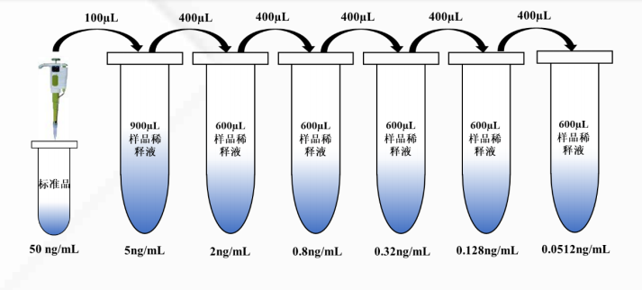 Restore the sample to room temperature and mix the sample before adding the sample; If the user needs to dilute the sample or the matching high-concentration standard in the box, the sample diluent in the box can be used for dilution; If it is a cell type sample, it is recommended to press 3000rpm/min Speed, centrifugal 5min Take the supernatant for detection. 2. Operation process: 1 Operation diagram 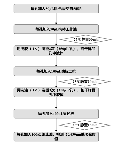 ( 1 ) Restore each component of the kit to room temperature 30min Take out the enzyme-labeled slats required for the test from the aluminum foil bag that has been equilibrated to room temperature, mark the order of the slats with a marker pen (it is recommended to perform double-well measurement), seal the remaining slats with a plate sealing film, put them back into the aluminum foil bag, seal them well, and place them in 2-8℃ Save. (Note: The slats are easy to fall off during the subsequent clapping process, so pay attention to marking them) ( 2 ) Sample incubation: Add to each well 50uL Standard / Blank (sample dilution) / Sample, after which added 50uL The antibody working solution was sealed with a plate sealing membrane and then placed in 25℃ Stationary incubation in the dark 30min 。 (Note: The standard must be added first. If the antibody is added first, it will react directly with the antigen on the plate; If the plate is not sealed or the plate is incompletely sealed during the incubation process, the reaction solution will evaporate, resulting in errors in the experiment; All incubation processes should be avoided as much as possible. Light irradiation) ( 3 ) Wash the plate: After the incubation is completed, carefully remove the plate sealing membrane, discard the liquid in the well, and use the washing solution ( 1× ) Wash the plate 3 Second ( 250uL/ well), and pat dry the residual liquid in the sample well. (Note: If hand washing board is used, add washing solution ( 1× ) When it is necessary to suspend liquid, it is best not to touch the inner wall of the hole; Each addition of the lotion ( 1× ) after resting 30s (under the last slight shake of the standing process); When patting dry, be careful to change the absorbent paper each time or pat dry on a clean area of the paper) ( 4 ) Enzyme-labeled secondary antibody incubation: enzyme-labeled secondary antibody is added to each well, 100uL/ Well, seal the plate with a plate sealing film, and then place it in 25℃ Stationary incubation in the dark 30min 。 ( 5 ) Wash the plate: the method is the same as the procedure ( 3 )。 ( 6 ) Color development: press the pre-configured color developing liquid 100uL/ The wells are added into the enzyme label plate, and the plate is sealed with a plate sealing membrane, 25℃ Stationary incubation in the dark 15min 。 ( 7 ) Termination: Adding a termination solution, 100uL/ Holes, which can be read after uniform color development. (Note: It is recommended to set in the microplate reader reading program 5-10s Oscillation of) ( 8 ) Reading: Put the enzyme label plate into the microplate reader and set the wavelength to double wavelength 450/630nm , read the absorbance value, the measurement should be after termination 20min Inside completion. III. Data processing: 1 Calculation of absorbance value The absorbance value of each standard or sample is OD450nm-OD630nm 。 2 Calculation of percent absorbance Mean absorbance value (double well) for each standard or sample divided by 0ng/mL The mean absorbance value of the standard is multiplied by 100% That is, the percent absorbance: Percentage absorbance ( % )= B/B0×100% B : Mean absorbance value of standard or sample B0 : 0ng/mL Mean absorbance values of standards 3 Drawing and calculation of standard curve Take the percentage absorbance value of the standard as the ordinate Y The standard concentration value is in the abscissa X , draw a standard curve, recommended using four parameters Logistic Mathematical model fitting equation: Y=((A-D)/(1+(X/C)^B))+D Substitute the percent absorbance value of the sample into the standard curve, read out the corresponding concentration value of the sample, and multiply it by its corresponding dilution factor to obtain the actual concentration of kanamycin in the sample.  |
||||||||||||||||||||||||||||||||||||||||||||
| Theory | This kit uses an indirect competitive ELISA method to determine trace residues of kanamycin in samples. The enzyme label plate is pre-coated with conjugated antigen, and the residual kanamycin in the sample and the conjugated antigen pre-coated on the microwell strip compete for anti-kanamycin antibody. The enzyme-labeled secondary antibody is added, and then the color is developed by adding TMB substrate. The absorbance (OD value) is measured at the wavelength of 450 nm/630 nm using a microplate reader, and the percent absorbance is calculated. The concentration of kanamycin in the sample is negatively correlated with the percent absorbance. | ||||||||||||||||||||||||||||||||||||||||||||
| Description | Kanamycin is an aminoglycoside antibiotic, which is used in the treatment of animal diseases, strain screening and preparation of drug raw materials for cell and gene therapy. 8 To the cranial nerves, resulting in vestibular and cochlear damage. The residues in animal food and biological drugs can affect human health and even cause allergic reactions. European and American countries and China all require its limited use. Kanamycin quantitative assay kit uses indirect competition ELISA Methods, the pre-coated conjugated kanamycin antigen on the microwell strip competes with the residual kanamycin in the sample to bind to the enzyme-labeled anti-kanamycin monoclonal antibody, and then the enzyme-labeled secondary antibody is added TMB The substrate developed color, and the absorbance value was measured using a microplate reader, which was negatively correlated with the content of kanamycin in the sample. The operation time of this kit is only about one hour, and the linear range is 0.05ng/mL-5ng/mL 。 When performing the linearity verification of kanamycin raw material, it is recommended that the kanamycin raw material be diluted to the linear range of the kit before determination, and diluted to at least a certain proportion 5 There was a linear correlation between the detection concentration and the dilution factor. Specificity should be validated for other substances present in the process. It is recommended to use high concentration and low concentration quality control samples and add increasing concentrations of related interfering substances for specificity investigation. The matrix without the addition of analytes should also be measured simultaneously. The accuracy of the controls should be within a certain range and the measurements of the matrix without the addition of analytes should be below the lower limit of quantification. Product Features: Strong specificity: No cross-reaction with penicillin, streptomycin sulfate, gentamicin and ampicin High Sensitivity: Kit sensitivity is 0.05ng/mL Convenient and efficient: Using a two-step method, only 1 Results can be obtained in about hours Excellent performance: good standard curve linearity, high accuracy and good repeatability Product performance indicators : Limit of detection: < 0.05 ng/mL Limit of Quantification: 0.05 ng/mL Linear Range: 0.05-5 ng/mL Accuracy (spiked recovery): 70%-130% Accuracy (measurement deviation): ≤15% Repeatability (intra-batch difference): ≤15% Specificity:
|
||||||||||||||||||||||||||||||||||||||||||||
| Composition |
|
||||||||||||||||||||||||||||||||||||||||||||
| General Notes | 1. The optimal reaction temperature of this kit is 25℃. If the temperature is too high or too low, the detection absorbance value and sensitivity will change. 2. All components in the kit must be restored to room temperature (20-25℃) before use. 3. Mix all components well before use, and the standard product needs to be briefly centrifuged for 5 seconds to concentrate all the liquid on the tube wall and cover at the bottom of the tube; Return all reagents to 2-8°C immediately after use. 4. The kit must be used within the shelf life, and the corresponding standard curve must be prepared for each test. It is not recommended to use different batches of relevant reagents in mixed batches. 5. Be careful not to touch the bottom of the microplate when adding liquid to the enzyme label plate to prevent damage to the coating layer. Replace the sampling tank and suction tip in time between different samples and steps to avoid cross-contamination. 6. When the slats are washed and dried, pay attention to prevent the slats from falling off, and the sealing film should not be reused. 7. High concentration may produce black flocs during color development, which is a normal phenomenon, and the slight degree does not affect the final reading result. 8. When reading, pay attention to whether the detection wavelength and fitting equation are selected correctly. 9. Only by strictly following the operation methods of the instructions and using all the reagents matched with this kit can the best detection effect be ensured. 10. The difference in test results can be caused by a variety of factors, including the operation of the experimenter, the use of the pipette, the washing technology, the reaction time or temperature, the storage of the kit, etc. 11. Our company is only responsible for the kit itself, not for the sample consumption caused by the use of the kit. Users are requested to fully consider the possible usage of samples before use and reserve sufficient samples. 12. The termination solution in this kit is an acid solution, so special attention should be paid to during operation. 13. Operators should wear personal protective equipment, such as laboratory coats, gloves, masks and goggles. |
||||||||||||||||||||||||||||||||||||||||||||
| Storage Temp. | The kit should be stored at 2-8℃, protected from light, with a shelf life of 12 months. Note that the unused kits after opening should still be stored at 2-8℃ in the dark. |