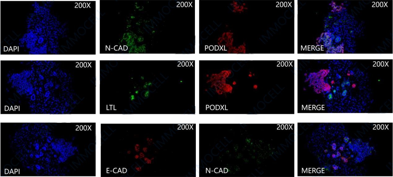
Immunofluorescence identification
hPSC Inducing Differentiation of Renal Organoids Kit
hPSC Inducing Differentiation of Renal Organoids Kit
Price:
Regular price
$4,794 USD
Regular price
Sale price
$4,794 USD
Unit price
per
For shipping services or bulk orders, you may request a quotation.
Secure checkout with

View full details
Product Details
Product Details
Product Specification
| Usage |
I. Experimental instruments and materials: hESC/iPSC a cell culture kit, hESC ( H1 )、 hESC/iPSC passage working fluid, Y-27632 、 DMEM/F12 Culture medium, growth factor reducing matrix glue, Accutase 、 PBS 、 12 well cell culture plates, 15 mL/50 mL centrifuge tube, 10 μL/200 μL/1000 μL a sterile tip, 10mL/50mL a pipette, DMSO 2. Reagent preparation:
2 In the biosafety cabinet, refer to the table. 1 and table 3 Using a sterile pipette and gun tip, mix well according to the proportions to prepare the culture medium for each stage of composition. 。 For example, induction medium 1 Composed of basic culture medium, 1× concentration of supplements A If the dosage is 50mL The preparation method is 49mL Basic medium +1mL supplements A ( 50× )。 3 Differentiation medium is recommended to be prepared and used immediately, and placed in 4℃ storage, 2 Use during the week. Note: All supplements can be repackaged according to the amount used to avoid repeated freezing and thawing. II. hESC Cultivation and preparation (see details) abs90487 ) III. DAY0-4: Late primitive stripe stage 1 Growth factor reduction matrix gel 4℃ Thawing below, with DMEM/F12 Homogeneous mixing (matrix glue :DMEM/F12=1:100 ), lay the boards, and set them aside. 2 、 DAY-1: When hESC The confluence of growth on the growth factor-reduced matrix gel reached approximately 40%-50% At left and right (density as shown in the figure below), aspirate the culture medium and add... 1mL Contains 1%DMSO pluripotent stem cell maintenance medium culture 24h 。
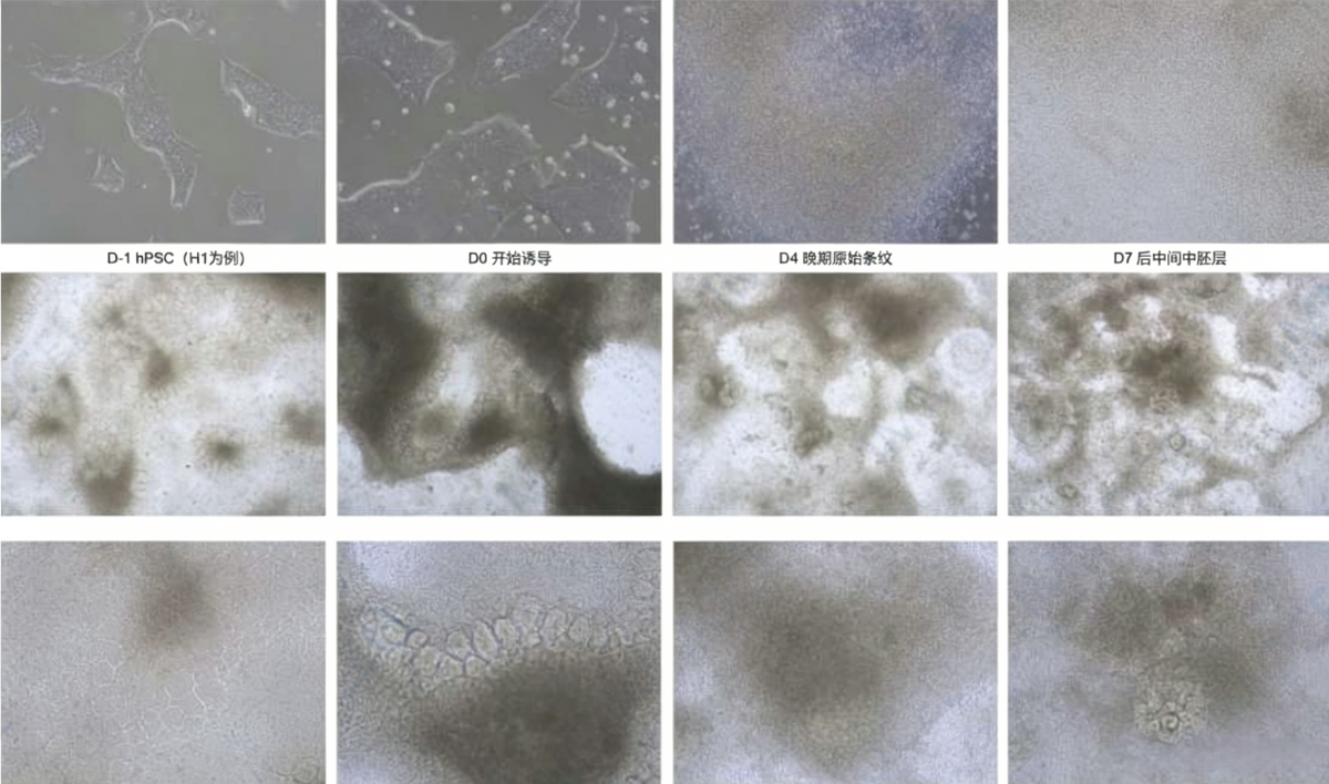
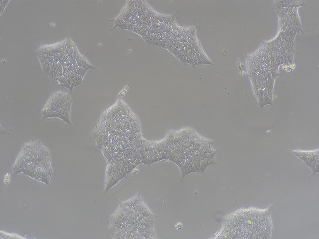 3 、 DAY0: Aspirate off the culture medium and add 1mL Induction medium 1 (The ingredients are: basic culture medium, 1× supplements A )。 3 、 DAY0: Aspirate off the culture medium and add 1mL Induction medium 1 (The ingredients are: basic culture medium, 1× supplements A )。 4 Use the induction medium every day or every other day depending on the color of the medium. 1 Change the fluid until the first 4 (Cell status as shown in the figure below). 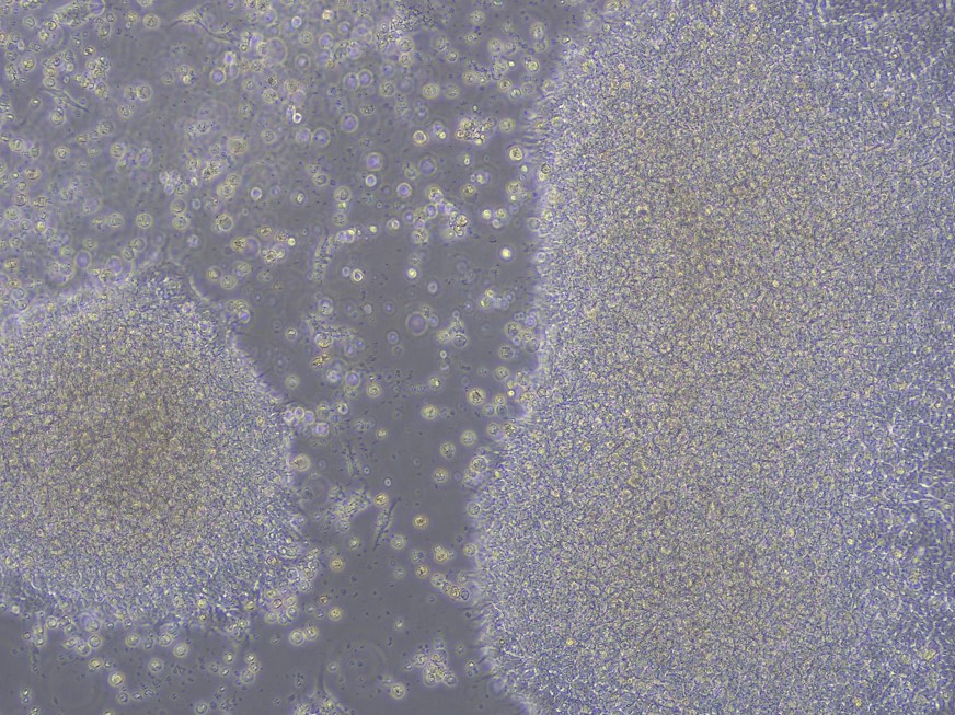 Note: ① When using the matrix adhesive, it must be handled on ice. All items in direct contact with the matrix adhesive must be pre-cooled beforehand. If the matrix adhesive exceeds... 10℃ It will solidify rapidly, resulting in failure of the plating. Note: ① When using the matrix adhesive, it must be handled on ice. All items in direct contact with the matrix adhesive must be pre-cooled beforehand. If the matrix adhesive exceeds... 10℃ It will solidify rapidly, resulting in failure of the plating. ② If the cell is in the first 3 A large number floated in the sky, or the first 3 There is a tendency for the edges to float and curl when changing the liquid every day; you can add... 10uM Y27632 Until the first 4 heaven. IV. DAY4-7: Post-intermediate mesoderm stage 1 、 DAY4 : The culture medium is aspirated from the culture, using 1mL Preheated Induction Medium 2 (The ingredients are: basic culture medium, 1× supplements B Change the fluid. 2 Use the induction medium every day or every other day depending on the color of the medium. 2 Change the fluid until the first 7 (Cell status as shown in the figure below). 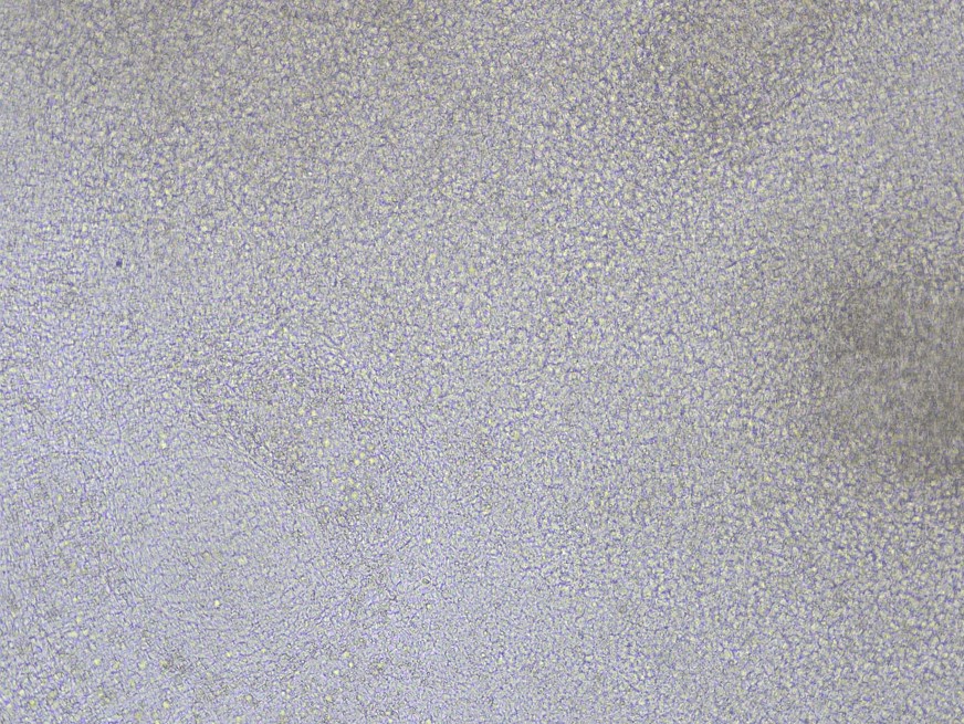 V. DAY7-9: Renal progenitor cell stage 1 、 DAY7: No 7 Days, the culture medium was aspirated from the culture, using 1mL Preheated Induction Medium 3 (The ingredients are: basic culture medium, 1× supplements C Change the fluid. 2 Change the medium every day or every other day depending on the color of the medium until the first... 9 (Cell status as shown in the figure below). 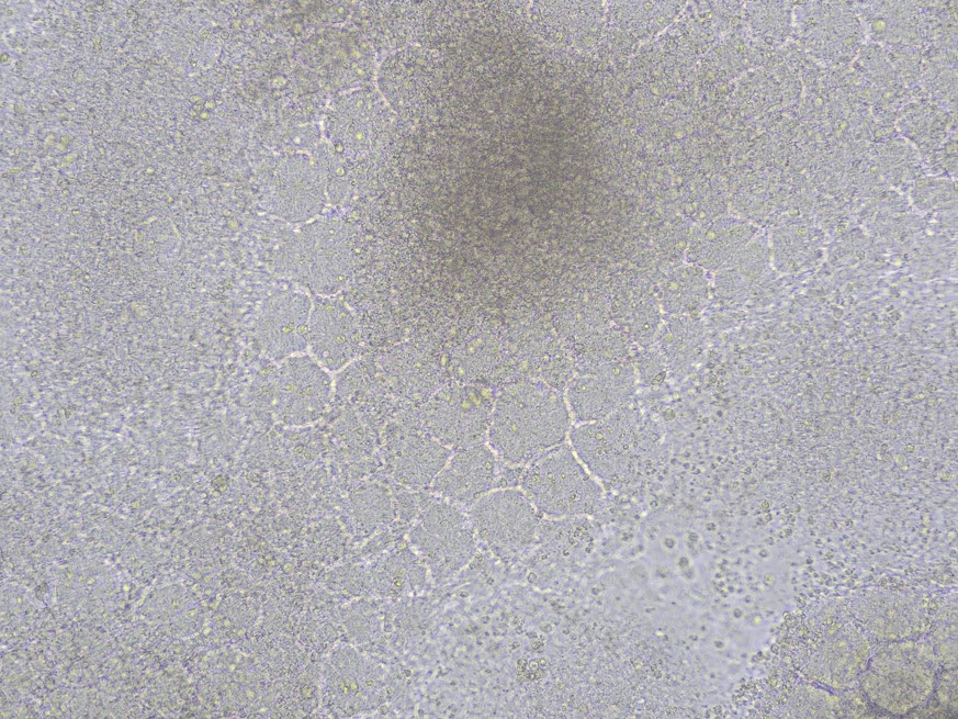 VI. DAY9-11: Pre-tubular aggregate stage 1 、 DAY9: The culture medium is aspirated from the culture, using 1mL Preheated Induction Medium 4 (The ingredients are: basic culture medium, 1× supplements D Change the fluid. 2 Change the medium every day or every other day depending on the color of the medium until the first... 11 (Cell status as shown in the figure below). 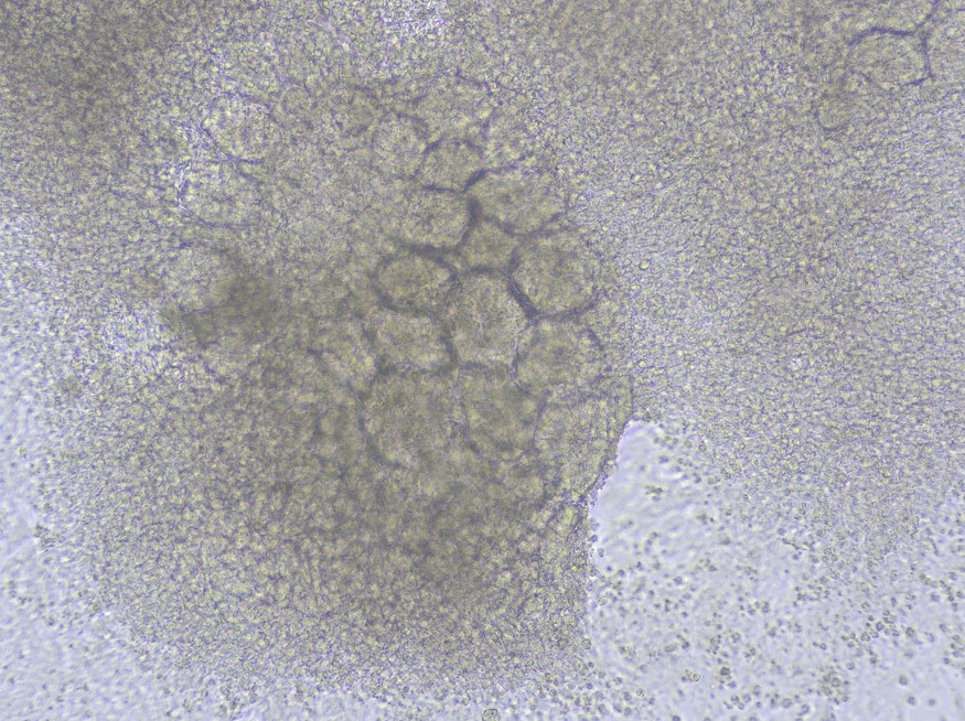 VII. DAY11-14: Pre-tubular aggregate stage 1 、 DAY11: The culture medium is aspirated from the culture, using 1mL Preheated Induction Medium 3 (The ingredients are: basic culture medium, 1× supplements C Change the fluid. 2 Change the medium every day or every other day depending on the color of the medium until the first... 14 (Cell status as shown in the figure below). 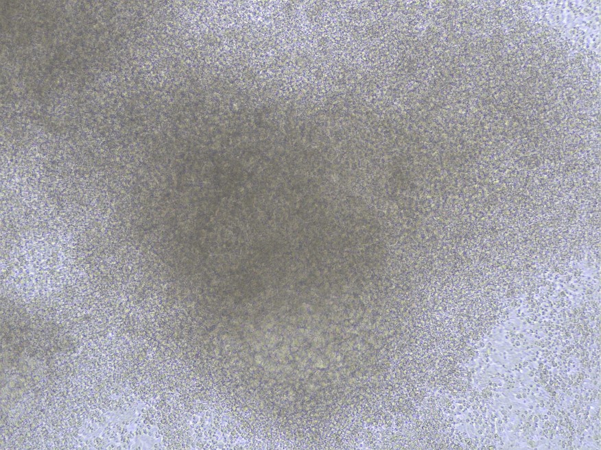 8. DAY14-21 or 40: Renal organoid maturation stage (1) DAY14: The culture medium is aspirated from the culture, using 1mL Change the preheated basal medium. (4) Daily or every other day depending on the color of the medium 1-2 Change the culture medium every day until the first day. 21 (Cell status as shown in the figure below). 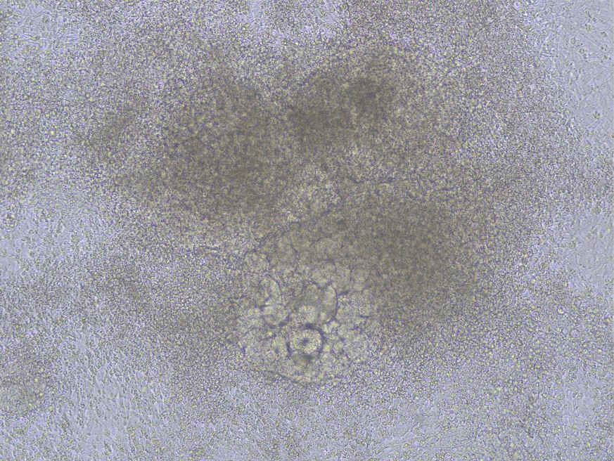 Note: No. 21 Renal organoid structures such as podocytes, proximal tubules, and distal tubules can be detected. For further maturation, they can be cultured until the first day. 40 heaven. Note: No. 21 Renal organoid structures such as podocytes, proximal tubules, and distal tubules can be detected. For further maturation, they can be cultured until the first day. 40 heaven. |
||||||||||||||||||||||||||
| Description | This kit is specially designed for inducing human pluripotent stem cells ( hPSC Designed for efficient differentiation into renal organoids, it provides a standardized, serum-free, and chemically defined culture system suitable for disease model construction, drug screening, and cell therapy research. The resulting renal organoids include podocytes, proximal tubules, and distal tubules, and can express specific markers (such as POXDL 、 LTL 、 E-CAD etc.), suitable for in vitro studies. With 12 Taking perforated plates as an example, the minimum specifications provided for this product are sufficient. 12 One hole hPSC Differentiation is induced, and based on cell state and cell line characteristics, each well can eventually obtain... 10-100 There are approximately six renal organoids. Product differentiation flow chart:  hPSC : human pluripotent stem cells hPSC : human pluripotent stem cells Product Composition:
|
||||||||||||||||||||||||||
| Application | This product requires operators to have cell culture experience and a certain understanding of long-term cell culture. | ||||||||||||||||||||||||||
| Storage Temp. | See product composition for details. |
Picture
Picture
Immunohistochemistry
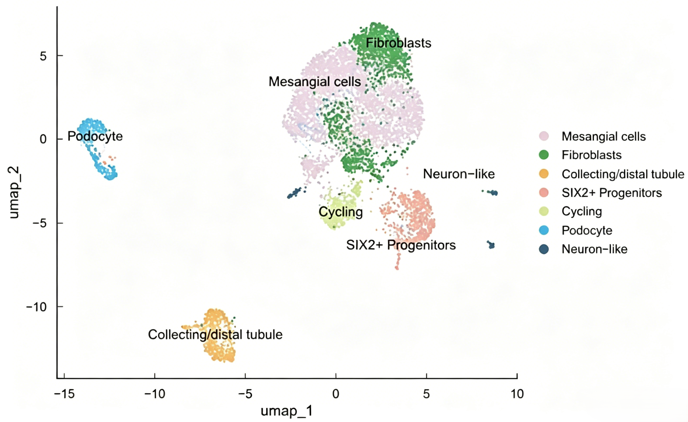
Based on the cell subpopulation types and characteristic marker genes of renal organoids, systematic cell annotations were performed on the single-cell sequencing clustering results, accurately identifying a variety of renal cell types with different developmental stages and functional phenotypes, fully demonstrating the core advantages of this model in highly replicating the physiological structure and embryonic development process of the human kidney. The model clearly identifies podocytes (podocytes, characteristically expressing NPHS1, NPHS2, WT1), Mesangial cells (Mesangial cells, highly expressing PDGFRB, ACTA2), and Fibroblasts (Fibroblasts, expressing COL1A1, VIM), and completely reduces the core structural cell components of the nephron. It is suitable for research on related mechanisms such as glomerular filtration and stromal microenvironment. At the same time, annotations to Collecting/distal tubule cells (expressing AQP2, SLC12A1) and SIX2-positive Progenitors (SIX2+ Progenitors, expressing SIX2, CITED1, PAX2) cover nephron epithelial differentiation and progenitor cell populations, which can support kidney development and regeneration research. In addition, Neuron-like cells and Cycling cells (expressing MKI67, TOP2A) were detected, suggesting that organoids have innervation simulation characteristics and self-renewal and proliferation capabilities, supporting long-term in vitro maturation and function maintenance. The overall cell population has good dispersion and clear subgroup boundaries, covering key types such as glomeruli, renal tubules, progenitor cells, stromal cells, and proliferating cells. It is highly close to the cellular composition and developmental characteristics of the human kidney, making it an ideal in vitro study of kidney development mechanisms, nephropathology, and drug screening. Model.
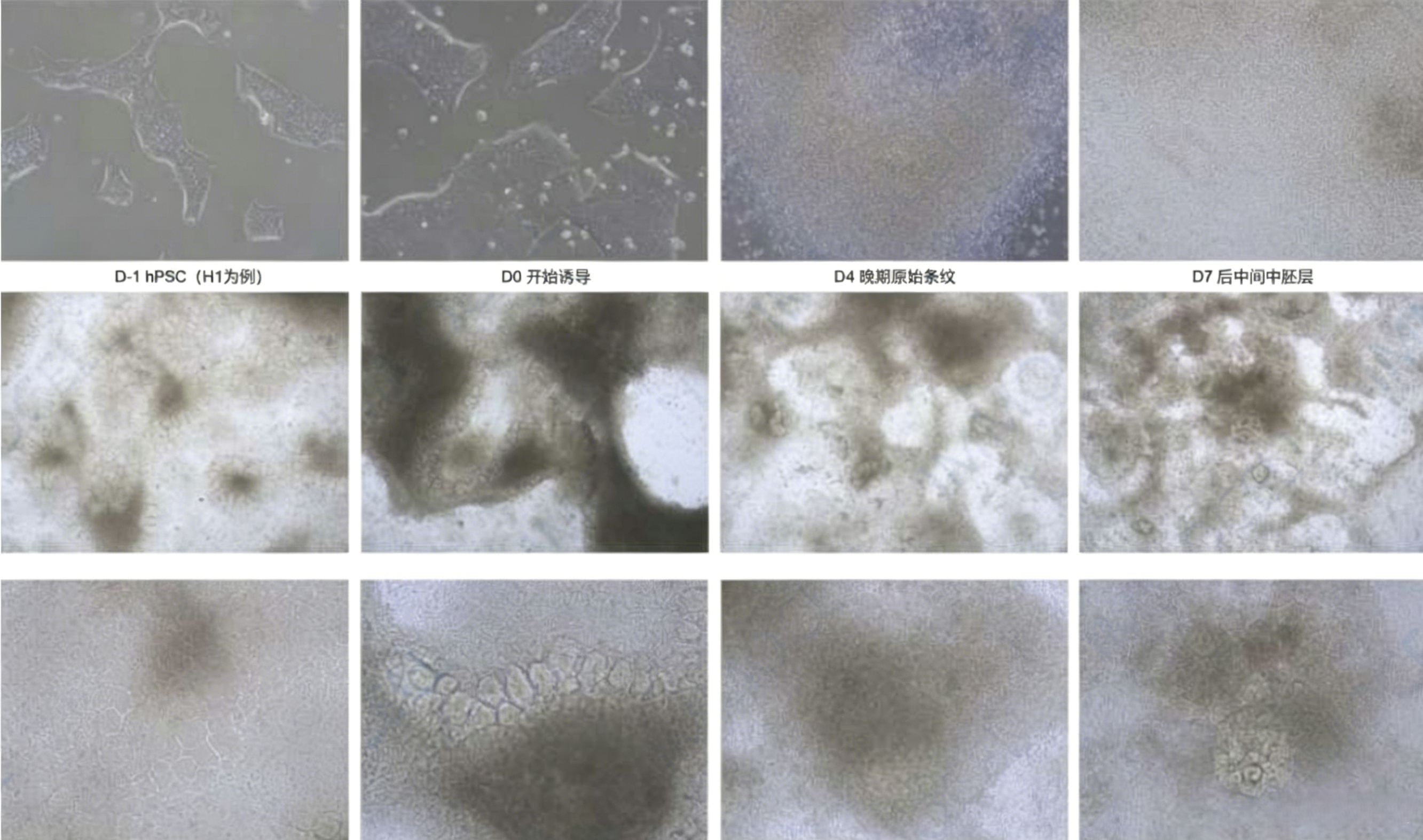
Cultured organoid images