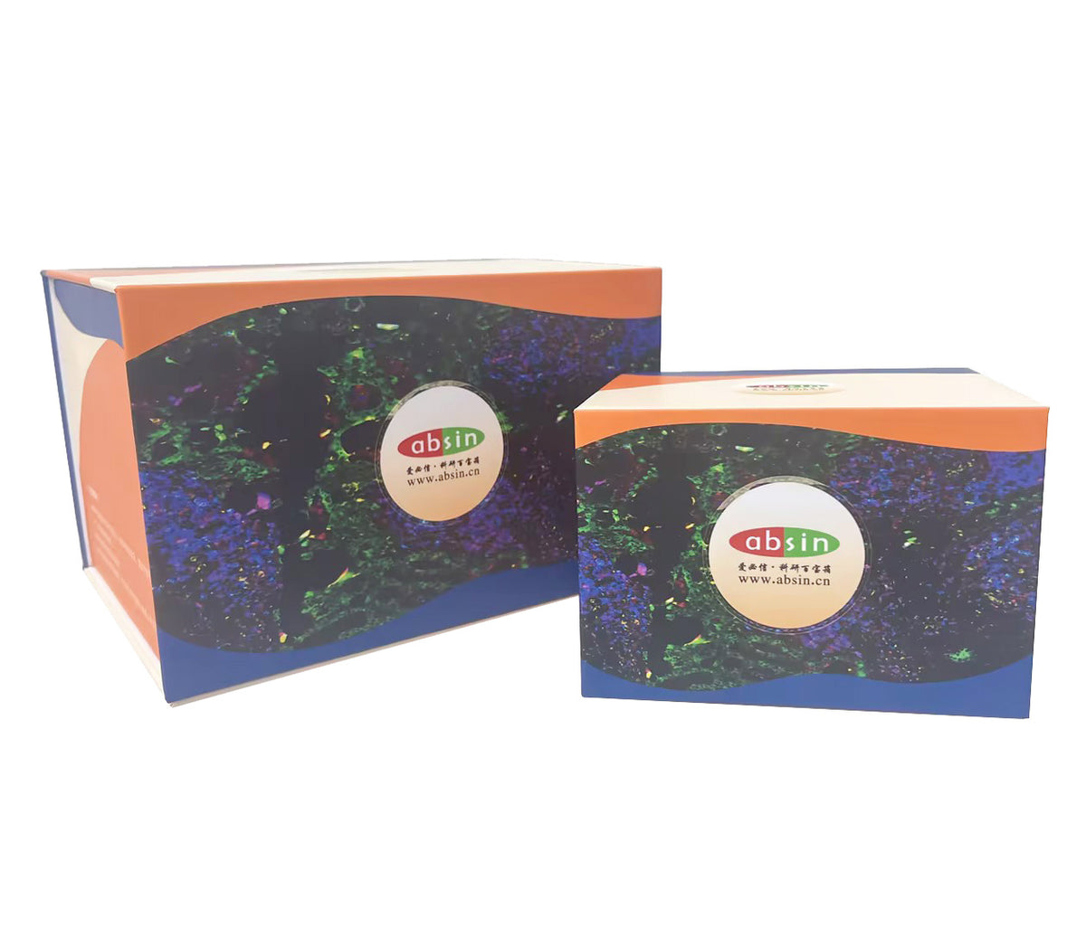
Absin 6-Color mlHC Kit(plus) (Anti-Rabbit Secondary Antibody)
Absin 6-Color mlHC Kit(plus) (Anti-Rabbit Secondary Antibody)

Product Details
Product Details
Product Specification
| References | 1. Mingzhu Yang, et al., Chemical-induced chromatin remodeling reprograms mouse ESCs to totipotent-like stem cells. Cell Stem Cell. 2022 Mar 3 2. Xin Guan, et al., Nanoparticle-enhanced radiotherapy synergizes with PD-L1 blockade to limit post-surgical cancer recurrence and metastasis. Nature Communication. 2022 May 20 3. Qian-Yue Zhang, et al., Lymphocyte infiltration and thyrocyte destruction are driven by stromal and immune cell components in Hashimoto’s thyroiditis. Nature Communications. 2022 Feb 9 4. Dabbs David J. 诊断免疫组织化学(M). 北京:北京大学医学出版社,2008.9. 5. 美】Ed Harlow, David Lane. 抗体技术指南(M). 北京:科学出版社,2002:79-80,105. 6. Stack, E. C., et al., Multiplexed immunohistochemistry, imaging, and quantitation: a review, with an assessment of Tyramide signal amplification, multispectral imaging and multiplex analysis. Methods, 2014 70 (1): 46-58. 7. 钱帮国. 焦磊. 多标记免疫荧光染色及多普成像技术在组织学研究中的应用. 中国组织化学与细胞化学杂志, 2017 (4): 373-382. |
| Usage | 1. For TSA dyes, please use signal amplification reaction solution diluted at 1:100 to prepare the dye working solution (currently used and prepared); DAPI prepared the working solution by diluting it with sterile water at a ratio of 1:100 |
| Description | There are complex cell composition in tissue microenvironment. The phenotype, state, abundance and distribution of these cells have important biological significance and clinical value. With the help of antibody staining, it can be presented in situ. Immunohistochemical staining is a common technology to study tissue morphology and in situ protein expression. Conventional IHC detection can only show a single index, which is difficult to show the cell composition, state and relationship in the complex tissue microenvironment, and this information is essential for the diagnosis and treatment of disease |
| General Notes | 1. This reagent kit is only used for immunohistochemistry and is not intended for any other purpose 2 This reagent kit is only for professional use 3 Appropriate protective measures should be taken to avoid contact between reagents and skin and eyes 4 Reagents that exceed their expiration date may have reduced activity, therefore they should not be used 5 If the staining components of this reagent kit are mixed with products from other companies, abnormal situations may occur during the staining process 6 Incomplete dewaxing can easily affect the dyeing effect 7 In order to prevent possible false negative and false positive results, both positive and negative controls should be conducted simultaneously during the experiment 8 All waste generated from the use of this reagent kit should be disposed of in accordance with the Medical Waste Management Regulations. |
| Storage Temp. | Fluorescent dyes need to be protected from light, Store at 2-8 ℃, 1 year from receipt |
| Applications | Used for immunohistochemical staining of tissues, paraffin sections, and TMA chips.Can also be used for frozen sections and cell slides with abs994 Antibody eluent (mIHC only) |
Picture
Picture
Immunohistochemistry