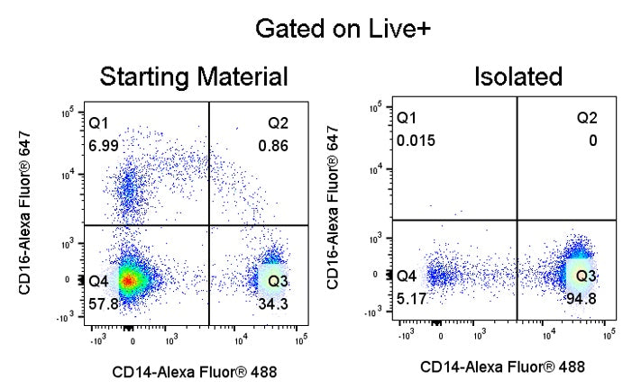
Human classical monocytes were enriched from peripheral blood mononuclear cells (PBMCs). After sorting, the cells were stained with CD14-Alexa Fluor® 488 (SDT Cat: S0B8022) and CD16-Alexa Fluor® 647 (SDT Cat: S0B85525), and analysis was performed by gating on live cells (Live+). Flow cytometric results showed that the purity of the sorted human classical monocytes (CD14+CD16-) was 94.8%.
Human Classical Monocyte Isolation Kit (Column-Based)
Human Classical Monocyte Isolation Kit (Column-Based)
Price:
Regular price
$1,335 USD
Regular price
Sale price
$1,335 USD
Unit price
per
For shipping services or bulk orders, you may request a quotation.
Secure checkout with

View full details
Product Details
Product Details
Product Specification
| Format |
1 mL Human Classical Monocyte Biotin-Antibody Cocktail; 0.2 mL Human FcR Blocking Reagent; 2 mL Streptavidin Beads; |
| Capacity | Applicable to1 × 10⁹Total cell count, capable of performing up to100sorts. |
| Principle of Separation | This reagentbox adoptsacolumn-based negative selectionmethod to isolateClassical Monocytesfrom peripheral blood. In this negative selection process, non-Classical Monocytecells are labeled with biotin-conjugated antibodies, and the cells are then incubated withstreptavidinaffinity nanomagnetic beads. Non-target cells are marked by the antibody complex binding to the magnetic beads, while the targetClassical Monocytecells remain unlabeled. Subsequently, the cell suspension is transferred to the separation column; under the influence of a magnetic field, the magnetically labeled non-target cells are adsorbed within the column, whereas the unlabeledClassical Monocytecells drip down into the collection tube under gravity. The resulting supernatant is the enriched fraction ofClassical Monocytecells after removal of non-target cells. |
| Applications in cell sorting | Classical monocytes in their native, unmanipulated state can be isolated from peripheral blood mononuclear cell (PBMC) samples and used for a variety of downstream functional assays. |
| Reactivity | Human |
| Magnetic Bead Size | 20 nm |
| Separation Method | Column-Based |
| Selection Strategy | Negative Selection |
| Stability & Storage | Store protected from light at 2–8 °C; do not freeze. |
Background
Human peripheral blood monocytes can be divided into three subpopulations: classical monocytes, non-classical monocytes, and intermediate monocytes. Among these, classical monocytes (CD14+CD16-) constitute the largest subset in peripheral blood. This population exhibits robust phagocytic activity and the capacity to secrete inflammatory cytokines, and under inflammatory microenvironments it can differentiate into functional cells such as macrophages and dendritic cells. As such, classical monocytes serve as a central cellular model for studying inflammatory mechanisms, immune regulation, cell differentiation pathways, and the development of therapeutics for inflammation‑related diseases. To obtain human classical monocytes in their native, unlabeled, and highly pure state, this kit employs a combination of specific antibodies to selectively label non‑target cells within peripheral blood mononuclear cells (PBMCs), including T cells, B cells, NK cells, granulocytes, platelets, and CD16‑negative non‑classical monocytes. These labeled cells are then removed via magnetic separation, leaving behind pristine classical monocytes that remain unmarked by any antibodies or magnetic beads. Compared with conventional sorting methods, the key advantage of this negative selection approach lies in its complete lack of labeling and direct contact with the target cells, thereby preserving the classical monocytes' original surface antigen profile, cellular viability, and biological functions to the greatest extent. The isolated cells can be directly used for immunological functional assays, induction of cell differentiation, investigations into inflammatory mechanisms, high‑throughput sequencing, in vitro drug screening, and basic immunological research, providing a stable and reliable cellular sample solution for all types of monocyte‑related studies.
Protocol
Note: This kit does not include the separation buffer; it must be provided separately. The corresponding Stater catalog number isS0D3018.
TakingL Separation Columnas an example
Steps |
Operating instructions |
Dosage and time |
|
Cell processing and labeling |
1 |
Isolate PBMCs from peripheral blood using density gradient centrifugation with lymphocyte separation medium, or directly thaw cryopreserved PBMC cells, and perform cell counting. Note: It is recommended to count cells after staining with AO/PI; PBMCs can be centrifuged at 400 g for 7 minutes. |
Sample preparation |
2 |
Resuspend 1×10in 100 μLMagSep Separation Buffer7Live cells; Note:Reagents should be adjusted proportionally according to the sample volume; set aside a pre-sorting sample to determine the proportion of Classical Monocytes; |
1×10⁷cells/100 μL |
|
3 |
Add 2 μL of FcR Blocking Reagent to the sample and gently mix; |
2 μL/100 μL |
|
4 |
Add 10 μL of Human Classical Monocyte Biotin Antibody Cocktail to the sample and gently mix; Note:For this step, gentle pipetting up and down 2–3 times can be used for mixing; |
10 μL/100 μL |
|
5 |
Incubate the antibodies in the cocktail with the cells at room temperature for 10 min; |
Incubate at room temperature for 10 min |
|
6 |
Mix the Streptavidin Beads well, then add 20 μL of Streptavidin Beads to the sample; |
20 μL/100 μL |
|
7 |
Gently mix the magnetic beads and cells, and incubate at room temperature for 5 minutes; Note:For this step, you can use a pipette to gently blow the mixture 2–3 times for mixing; |
Incubate at room temperature for 5 minutes |
|
8 |
Add a volume of MagSep Separation Buffer equal to 10 times the cell volumeWash the cells, centrifuge at 400g for 7 minutes, discard the supernatant, and resuspend to the original volume. |
Centrifuge at 400g for 7 minutes. |
|
Cell sorting |
nine |
In the column‑separation magnet, install the L separation column and add 3 mL.MagSep Separation Buffer, flush and clean the sorting column; |
Washing and sorting column |
10 |
Add the cells to the sorting column, and collect the target cells using a collection tube. Note:The cell volume shall be at least 1 mL; if insufficient, useMagSep Separation BufferComplete the process; cells that drip under gravity are the target cells. |
Collect target cells |
|
11 |
Add in three portions, 3 mL each.MagSep Separation Buffer, collect the target cells; Note:In this step, unlabeled target cells are eluted from the separation column to enhance the yield. |
Collect target cells |
|
Notes: Pay attention to the maximum load capacity of the separation column; for the S Separation Column, the maximum cell count is 1 × 10.seven; The maximum number of labeled cells in the L separation column is 1 × 10.8; | |||
Picture
Picture
Validation Data