Human CCL17 Kit (HICA)
Human CCL17 Kit (HICA)

Product Details
Product Details
Product Specification
| Stability & Storage | 2~8℃ Store away from light.6months; after reconstituting the calibrator, it can be aliquoted and stored at-20℃ for three months, avoiding repeated freeze-thaw cycles. |
Background
Human chemokine ligand17 (Human C-C motif chemokine 17, CCL17), also known as thymus and activation-regulated chemokine (TARC), is a small secreted protein encoded by the CCL17 gene on chromosome 16q21, with a molecular weight of approximately 8 kDa. It belongs to the CC family of chemokines; its molecule maintains a typical three-dimensional folded structure stabilized by two disulfide bonds and can spontaneously form non-covalent dimers or polymers at high concentrations. CCL17 is constitutively expressed in immune organs such as the thymus and lymph nodes, and can be produced by mature dendritic cells, Langerhans cells, monocytes/macrophages,endothelial cells, keratinocytes, and airway epithelial cells inCCL17 is highly induced by inflammatory signals such as IL-4, IL-13, and TNF-α, and serves as a key chemokine within the Th2-type immune microenvironment. It primarily exerts its function by binding with high affinity to CCR4, selectively recruiting CCR4-expressing Th2 cells, regulatory T cells (Tregs), skin-homing memory T cells, basophils, and iNKT cells, thereby playing a central role in the directional migration of immune cells, tissue homing, and the formation of lymphoid organ architecture. Additionally, CCL17 can interact with CCR2 and the atypical chemokine receptor ACKR2 (D6), contributing to the fine-tuned regulation of the chemokine network. Under pathological conditions, the CCL17/CCR4 pathway…The shaft is the drive.One of the core pathways of Th2 inflammatory responses, it can promote the recruitment of Th2 cells to inflammatory sites, induce basophil activation and IgE class switching, and synergize with cytokines such as IL-4, IL-5, and IL-13 to amplify allergic reactions. In patients with atopic dermatitis, serum TARC levels are highly correlated with disease severity and rash area, making it a commonly used biomarker for clinically assessing Th2 inflammatory activity. Additionally, CCL17 is significantly elevated in other type 2 inflammatory diseases, including allergic asthma, allergic rhinitis, eosinophilic esophagitis, and chronic sinusitis with nasal polyps. In the field of oncology, CCL17 can be produced by Reed-Sternberg cells in Hodgkin lymphoma, as well as by adult T-cell leukemia/lymphoma (ATLL) and mycosis fungoides.etc.CCR4⁺Tumor cells exhibit high CCL17 expression, which recruits Tregs to establish an immunosuppressive microenvironment and is closely associated with disease progression and prognosis. Given CCL17's pivotal role in type 2 inflammatory diseases, allergic skin disorders, and lymphatic system malignancies, it has garnered significant attention in studies of disease mechanisms, therapeutic efficacy monitoring, and biomarker development. Researchers frequently assess CCL17 levels to evaluate the strength of Th2 immune responses, the activity of allergic diseases, and the effectiveness of interventions targeting IL‑4Rα or CCR4. The Human CCL17 ELISA Kit provides a sensitive, specific, and efficient tool for the quantitative in vitro detection of CCL17 concentrations in cell culture supernatants, serum, plasma, and other biological samples, making it suitable for basic biomedical research and drug screening applications.
Principle of the test:
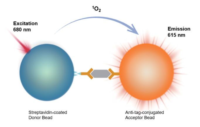
This kit employs a homogeneous chemiluminescence assay based on the sandwich ELISA method.Homogeneous Immuno Chemiluminescence Assay, HICA) to detect cytokine concentrations, with simple operation,No washing required.
The detection system contains two types of microspheres:Receptor microspheres are conjugated with antibodies targeting the protein of interest.1. Donor microspheres are conjugated with streptavidin, while antibody 2 is labeled with biotin. During the reaction, the target protein binds to the antibody and the microspheres, forming an immune complex that brings the two types of microspheres into close proximity. When the distance between the donor and receptor microspheres is less than 200 nm, singlet oxygen generated upon light excitation can be transferred to the receptor microsphere, triggering chemiluminescence; conversely, if the target protein is absent and the microspheres are too far apart, no signal is produced.
By measuring the intensity of the chemiluminescence signal, quantitative analysis of the target protein can be achieved. This method is characterized by its simplicity, rapidity, and high sensitivity.
Components
Name |
Specification |
Component Specification |
Test Reagent R1 |
100 tests |
2.4 μl/tube × 1 |
500 tests |
12 μl/tube × 1 |
|
Test Reagent R2 |
100 tests |
480 μl/tube × 1 |
500 tests |
2.4 ml/tube × 1 |
|
Test Reagent R3 |
100 tests |
6 μl/tube |
500 tests |
30 μl/tube |
|
Calibrator |
100 tests |
30 ng lyophilized powder × 1 |
500 tests |
30 ng lyophilized powder × 5 |
|
Reagent Diluent |
100 tests |
1.6 mL/bottle × 1 |
500 tests |
8 mL/bottle × 1 |
|
Calibrator Diluent |
100 tests |
1 mL/tube × 1 |
500 tests |
1 mL/tube × 5 |
|
RPMI Diluent |
100 tests |
400 μl/tube |
500 tests |
1 mL/tube × 2 |
Note: It is recommended to use a microplate for the plates used.384Shallow hole or 384,96Orifice plate, white or gray)
Protocol
I. Sample Requirements
1.To remove impurities from the sample, the sample must be centrifuged prior to analysis.2000 × g,10minutes).
2.If the measured concentration of a sample exceeds the highest value on the standard curve, it should be appropriately diluted (using negative serum is recommended) and retested to ensure that the result falls within the valid measurement range.
II. Testing Procedure
2.1Preparation of calibration standard gradient samplesAdopt 100muLReconstitute with deionized water.30Lyophilized calibrator, then dilute the reconstituted lyophilized calibrator with calibrator diluent; calibrator C.1This is the calibrator diluent; the recommended dilution protocol is shown in the table below:
Gradient |
Concentration (pg/mL) |
Higher-level calibrator(microliters) |
Diluent (μL) |
C11 |
100,000 |
30 μLReconstituted calibrator |
60 |
C10 |
30,000 |
30μL C11 |
70 |
C9 |
10000 |
30μL C10 |
60 |
C8 |
3000 |
30μL C19 |
70 |
C7 |
1000 |
30μL C8 |
60 |
C6 |
300 |
30μL C7 |
70 |
C5 |
100 |
30μL C6 |
60 |
C4 |
30 |
30μL C5 |
70 |
C3 |
10 |
30μL C4 |
60 |
C2 |
3 |
30μL C3 |
70 |
C1 |
0 |
- |
- |
2.2Testing procedure:
|
Testing procedure 25°C test | |
First timeSample addition |
1.Take2µL sampleAdd to384microplate; 2.PutAfter sonicating R1 for 5 seconds or vortexing, dilute it 200-fold with the reagent diluent; then add an equal volume of vortexed R2 and mix thoroughly to obtain the pre-mixed R1/R2 working solution (R1∶R2 = 1∶1). It is recommended to use 8 per well.muPrepare L × (number of reaction wells + 10%); 3.Add to each well8 μL of premix working solution. |
First incubation |
In a microplate shaker800 r/min 25degreeCIncubation60min |
Second sample addition |
Before the first incubation endsIncubate for 5–10 minutes, sonicate R3 for 5 seconds or vortex to mix thoroughly, and dilute with the reagent diluent by a factor of 200; add 10 μL of the R3 working solution to each well. |
Second incubation |
800 r/min,25degreeC. Incubate in the dark for 30 min. |
Reading |
Instrument reading,Shade from light/Green light |
* Note:Before use, be sure to sonicate R1 and R3 for 5 seconds or vortex them thoroughly., prepare and use immediately, requiredShade from light/Green light
When adding samples, keep the pipette tip close to the bottom of the well to avoid introducing air bubbles, as bubbles can affect the readings.
The reaction system can be scaled proportionally according to experimental requirements.
III. Performance Testing
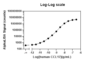
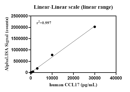
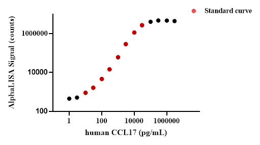
3.1Example of a complete standard curve:
3.2Performance Parameter Verification:
LOB:Repeat detection20Next calibratorC1, calculate the signal mean andSD, calculate using the standard curveSignal mean+2timesSDThe corresponding concentration value is the limit of detection (LOD).B.
Matrix |
Blank limit (pg/mL) |
Buffer solution |
2.3 |
Dynamic rangeDynamic range):0 –100 000 pg/mL.
Linear range (Linear Range):10 – 30,000 pg/mL.
Precision (Precision):
Intra-precision (Intra-Precision): In the same experimentOn the orifice plate, using known low,China,Each high-concentration sample was tested in triplicate.1Zero replicates were performed to evaluate the intra-assay precision of the assay. The coefficient of variation (%CV) for each concentration was less than 10%, indicating that the method exhibits good reproducibility within the same batch..
Matrix |
Precision |
||
Low concentration |
Medium concentration |
high |
|
Buffer solution |
7% |
2% |
3% |
Inter-plate precision (Inter-Precision): In 5In the next independent experiment, known low levels were tested separately,China,High-concentration samples are tested. Settings5The results for samples at each concentration exhibited low coefficients of variation (CV < 10%), indicating that the method demonstrates good reproducibility across different assay batches.
Matrix |
Intermediate precision |
||
Low concentration |
Medium concentration |
High concentration |
|
Buffer solution |
9% |
4% |
3% |
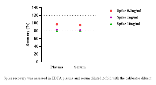
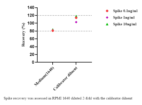
Accuracy (Recovery):
Separately, in different matrices (the blood matrix was diluted 2-fold with the calibrator diluent, and the culture supernatant was used...RPMIDiluentAdd low, medium, and high concentrations of C to the 2-fold diluted solution.CL17For the standard, perform duplicate tests to determine its average recovery rate.
Matrix |
Recovery rate |
||
Low concentration |
Medium concentration |
High concentration |
|
EDTAPlasma |
97% |
83% |
80% |
serum |
95% |
82% |
81% |
Culture medium supernatant |
81% |
82% |
84% |
Calibrator diluent |
114% |
10.03% |
118% |
Dilution linearity (Dilution linearity):
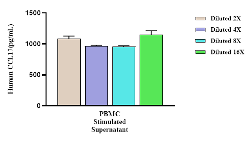
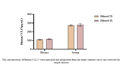
Separately for PBMC(10 μg/mL PHA stimulationPeripheral blood mononuclear cells72 hoursConduct gradient dilutions and perform calibration, as well as conduct gradient dilutions and calibration for human serum and plasma, to evaluate their dilution linearity.
Hook effect (HOOK):
takeCCL17Standard substanceGradient dilution and testing confirmed that samples within the measurement range yield reliable quantitative results, and no signal suppression was observed.
Guidelines