
Human FVIIa ELISA Kit
Human FVIIa ELISA Kit
Price:
Regular price
$368 USD
Regular price
Sale price
$368 USD
Unit price
per
For shipping services or bulk orders, you may request a quotation.
Secure checkout with

View full details
Product Details
Product Details
Product Specification
| Usage |
1. Self-prepared test equipment required for the experiment: 1 , plate reader ( 450nm ) 2 , high-precision sampler and gun head: 0.5-10μL 、 5-50μL 、 20-200μL 、 200-1000μL 3 , 37℃ Incubator 4, Distilled water or deionized water, 2. Sample processing and requirements: 1, Serum: the whole blood sample collected in the serum separation tube is placed at room temperature 2 Hour or 4℃ Overnight, then 1000×g Centrifugation 20 Minutes, take the supernatant, or place the supernatant in -20℃ Or -80℃ Store, but repeated freezing and thawing should be avoided. 2, Plasma: with EDTA Or heparin as an anticoagulant to collect specimens, and collect the specimens after collection 30 Within minutes 2-8℃ 1000 ×g Centrifugation 15 Minutes, take the supernatant to detect, or place the supernatant in -20℃ Or -80℃ Store, but repeated freezing and thawing should be avoided. 3, Tissue homogenate: with pre-cooled PBS (0.01M, pH=7.4) The tissue is flushed to remove residual blood (lysed red blood cells in the homogenate can affect the measurement), and the tissue is weighed and cut into pieces. Combining the shredded tissue with the corresponding volume PBS (Generally according to 1:9 Weight to volume ratio, such as 1g The tissue samples correspond to 9mL Of PBS The specific volume can be appropriately adjusted according to the experimental needs and recorded. Recommended in PBS Add protease inhibitor) into a glass homogenizer and ground thoroughly on ice. For further lysis of tissue cells, the homogenate can be sonicated, or freeze-thawed repeatedly. Finally, the homogenate was mixed in 5000×g Centrifugation 5~10 Minutes, take the supernatant for detection. 4 , cell culture supernatant or other biological specimen: please 1000×g Centrifugation 20 Minutes, take the supernatant to detect, or place the supernatant in -20℃ Or -80℃ Store, but repeated freezing and thawing should be avoided. Note: Hemolysis of the specimen will affect the final test result, so hemolyzed specimens should not be tested. 3. Preparation work before testing: 1, The kit should be taken out of the refrigerated environment and equilibrated at room temperature before use. 2 , 20× Dilution of wash buffer: distilled water according to 1 : 20 Dilution, i.e. 1 Share 20× Wash buffer plus 19 Distilled water. 4. Operation steps: 1, Equilibration from room temperature 20min Take out the required slats from the aluminum foil bag after that, and seal the remaining slats with ziplock bag and put them back 4℃ 。 2, Standard hole and sample hole are set, and different concentrations of standards are added to each standard hole 50μL ; 3, The sample to be tested is added into the sample hole 50μL ; Blank holes are not added. 4, Except for the blank wells, horseradish peroxidase ( HRP ) Labeled detection antibody 100μL Sealing the reaction hole with a sealing plate membrane, 37℃ Incubation in water bath or incubator 60min 。 5, discard the liquid, pat dry on absorbent paper, and fill each well with washing liquid ( 350μL ), let stand 1min , throw off the washing liquid, pat dry on absorbent paper, and repeat washing the plate 5 Times (the plate can also be washed with a plate washing machine). 6, Adding substrate per well A 、 B Each 50μL , 37℃ Incubate in the dark 15min 。 7, adding stop solution to each well 50μL , 15min Inside, in 450nm Wavelength measurement of each well OD Value. 5. Calculation of experimental results: Result judgment: Based on the tested standard OD The value is the abscissa, and the concentration value of the standard product is the ordinate. Draw the standard curve on coordinate paper or with relevant software, and obtain the linear regression equation. The sample OD Values are substituted into Eq., and the concentration of the sample is calculated. 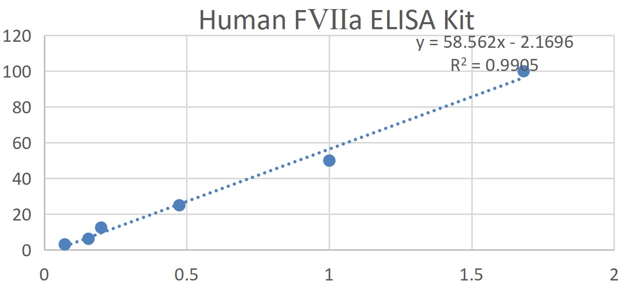 6. Kit performance 1, Detection range: 3.125 ng/ml – 100 ng/ml 。 2, Sensitivity: the lowest detection concentration is less than 0.1 ng/ml 。 3, Specificity: does not cross-react with other soluble structural analogs. 4, Repeatability: the coefficient of variation in the plate is less than 10% , the interplate coefficient of variation is less than 15% 。 7. Description Due to the existing conditions and scientific and technological level, it is not possible to comprehensively identify and analyze all raw materials provided by all suppliers, and this product may have certain quality and technical risks. 1, The final experimental results are closely related to the effectiveness of the reagents, the relevant operations of the experimenter and the experimental environment at that time. Please be sure to prepare sufficient specimen backups. 2, Different batches of the same product may have slight differences, such as detection limit, sensitivity and color development time, etc. Please conduct experimental operations according to the instructions in the kit. The electronic version of the instructions on the website is for reference only. 3, Only by using all the supporting reagents of this kit can the detection effect be guaranteed, and products of other manufacturers cannot be mixed. The best test results will only be obtained if the experimental instructions of this kit are strictly followed. 4, The company is only responsible for the kit itself and is not responsible for the sample consumption caused by the use of the kit. Users are requested to fully consider the possible usage of samples and reserve sufficient samples before use. 5, Tissue homogenates or cell extracts prepared using chemical lysates may be caused by the introduction of certain chemicals ELISA Experimental results bias. 6, If the sample is cell culture supernatant, there may be cases where it cannot be detected because there are many interference factors in this type of sample, such as cell status, cell number, sampling time, etc. 7, Certain natural proteins or recombinant proteins, including prokaryotic and eukaryotic recombinant proteins, may not be detected because they do not match the detection antibodies and capture antibodies used in this product. |
|||||||||||||||||||||||||||||||||
| Sensitivity | Minimum detected concentration less than 0.1 ng/ml | |||||||||||||||||||||||||||||||||
| Theory | The kit uses a double-antibody one-step sandwich enzyme-linked immunosorbent assay (ELISA). To the coated microwells pre-coated with human coagulation factor Ⅶ a (F Ⅶ a) capture antibody, specimens, standards, and HRP-labeled detection antibodies were added sequentially, incubated and washed thoroughly. The color is developed with the substrate TMB, which is converted to blue under the catalysis of peroxidase and to the final yellow under the action of acid. The depth of color was positively correlated with human coagulation factor Ⅶ a (F Ⅶ a) in the sample. The absorbance (OD value) was measured with a microplate reader at a wavelength of 450nm, and the sample concentration was calculated. | |||||||||||||||||||||||||||||||||
| Source | Human | |||||||||||||||||||||||||||||||||
| Synonym | Human Activated Coagulation Factor VIIa ELISA Kit | |||||||||||||||||||||||||||||||||
| Detection Type | Double antibody sandwich method | |||||||||||||||||||||||||||||||||
| Composition |
1. The standard concentrations are as follows: 100 、 50 、 25 、 12.5 、 6.25 、 3.125 ng/mL 。 |
|||||||||||||||||||||||||||||||||
| Background | Factor VII (formerly known as proconvertin) is one of the proteins in the clotting cascade that causes blood to clot. It is an enzyme of the class of serine proteases. Once bound to tissue factor released from damaged tissue, it is converted to factor VIIa (or hemagglutination factor VIIa, activated hemagglutination factor VII), which in turn activates factors IX and X. | |||||||||||||||||||||||||||||||||
| General Notes | 1. Carry out incubation in strict accordance with the specified time and temperature to ensure accurate results. All reagents must reach room temperature 20-25 °C prior to use. Store reagents in refrigeration immediately after use. 2. Incorrect plate washing can lead to inaccurate results. Make sure to drain the liquid from the wells as much as possible before adding the substrate. Do not allow the wells to dry out during incubation. 3. Eliminate the residual liquid and fingerprints at the bottom of the plate, otherwise it will affect the OD value. 4. The substrate color development solution should be colorless or very light in color, and the substrate solution that has turned blue cannot be used. 5. Avoid cross-contamination of reagents and specimens to avoid wrong results. 6. Avoid direct exposure to strong light during storage and incubation. 7. After balancing to room temperature, open the sealed bag to prevent water droplets from condensing on the cold slats. 8. Any reaction reagent cannot come into contact with the bleaching solvent or the strong gas emitted by the bleaching solvent. Any bleaching component will destroy the biological activity of the reaction reagents in the kit. 9. Expired products cannot be used. 10. If the disease may be spread, all samples should be managed well, and the samples and testing devices should be handled according to the prescribed procedures. |
|||||||||||||||||||||||||||||||||
| Storage Temp. | Storage conditions: 2-8 ℃, shelf life: 6 months. | |||||||||||||||||||||||||||||||||
| Test Range | 3.125 ng/ml – 100 ng/ml |