Standard curve
Example of MCP-1 (CCL2) standard curve in Assay Diluent .
Human MCP-1 (CCL2) Single Plex Kit (Flow Cytometry Bead Assay)
Human MCP-1 (CCL2) Single Plex Kit (Flow Cytometry Bead Assay)
Price:
Regular price
$500 USD
Regular price
Sale price
$500 USD
Unit price
per
For shipping services or bulk orders, you may request a quotation.
Secure checkout with

View full details
Product Details
Product Details
Product Specification
| Antigen | MCP-1 (CCL2) |
| Reactivity | Human |
| Stability & Storage | 12 months from date of receipt / reconstitution, 2 to 8°C as supplied. |
Kit
| Precision | Intra-assay: 3.19%; Inter-assay: 2.04% |
| Sample type | Serum; Plasma-EDTA; Plasma-Heparin; Plasma-Citrate; Cell Culture Supernatant |
| Assay type | Sandwich (quantitative) |
| Sensitivity | 1.77 pg/mL |
| Range | 2.03 pg/mL-8300 pg/mL |
| Recovery | Serum: 90.72% Plasma-EDTA:102.64% Plasma-Heparin:83.02% Plasma-Citrate:110.05% Cell culture supernatant: 124.14% |
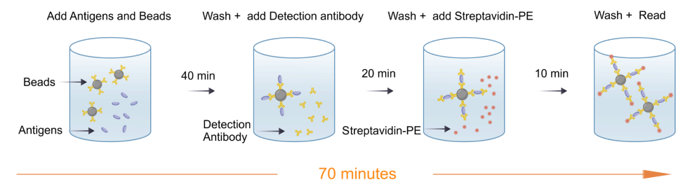
| Assay time | 70 minutes |
| Species reactivity | Human |
Background
MCP-1, also known as CCL2, is a chemokine belonging to the C-C chemokine family. It plays a crucial role in various physiological processes such as inflammation, immune regulation, and cell migration. MCP-1/CCL2 primarily functions by binding to the C-C chemokine receptor CCR2, thereby guiding monocytes and other inflammatory cells to migrate to sites of inflammation, contributing to the inflammatory response and immune regulation. Overexpression of MCP-1/CCL2 is closely associated with the pathogenesis and progression of various diseases, including inflammatory diseases, autoimmune disorders, cancer, and metabolic disorders. As a result, MCP-1/CCL2 has emerged as a potential therapeutic target, and researchers are exploring therapeutic strategies targeting MCP-1/CCL2 in the hope of impacting the treatment of related diseases.
Picture
Picture
CBA
Quantifiction of human PBMC
Stimulated sample: Human peripheral blood mononuclear cells (PBMCs) were stimulated with 2 µg/mL PMA for 22h, and the average concentration of MCP-1 (CCL2) in the supernatant was measured to be 9826.70 pg/mL.
Unstimulated sample: Human PBMCs were cultured for 22h, and the average concentration of MCP-1 (CCL2) in the supernatant was measured to be 82.20 pg/mL.
Linearity
The high-concentration human MCP-1 (CCL2) protein was serially diluted using serum, plasma-EDTA、 plasma-heparin, plasma-citrate and cell culture supernatant matrices respectively. Then, the concentration of human MCP-1 (CCL2) was determined and interpolated based on the target standard curve, and the sample dilution was corrected.
Protocol Diagram