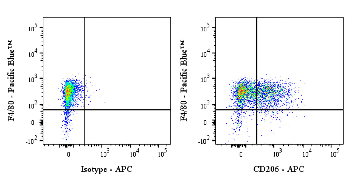
Flow cytometric analysis of CD206 (MMR) expression on 4% paraformaldehyde fixed and 0.1% Tween permeabilized BALB/c mouse peritoneal exudates cells. The cells were then stained with PE Rat Anti-Mouse F4/80 Antibody and either APC Rat IgG2a, κ Isotype Control (left panel) or SDT APC Rat Anti-Mouse CD206 (MMR) Antibody (right panel) at 1.25 μl/test. Total viable cells, as determined by Fixable Viability Dye 545 (S0B88802), were used for analysis. Flow cytometry and data analysis were performed using Agilent NovoCyte Quanteon and FlowJo™ software.
APC Rat Anti-Mouse CD206 (MMR)Antibody (S-R498)
APC Rat Anti-Mouse CD206 (MMR)Antibody (S-R498)
Price:
Regular price
$150 USD
Regular price
Sale price
$150 USD
Unit price
per
For shipping services or bulk orders, you may request a quotation.
Secure checkout with

View full details
Product Details
Product Details
Product Specification
| Host | Rat |
| Antigen | CD206 (MMR) |
| Location | Cell membrane |
| Accession | Q61830 |
| Clone Number | S-R498 |
| Antibody Type | Rat mAb |
| Isotype | IgG2a,k |
| Application | ICFCM, FCM |
| Reactivity | Ms |
| Positive Sample | BALB/c mouse peritoneal exudates cells |
| Purification | Protein G |
| Concentration | 0.2 mg/ml |
| Conjugation | APC |
| Physical Appearance | Liquid |
| Storage Buffer | PBS, 1% BSA, 0.3% Proclin 300 |
| Stability & Storage | 12 months from date of receipt / reconstitution, 2 to 8 °C as supplied |
Dilution
| application | dilution | species |
| ICFCM | 0.25μg per million cells in 100μl volume | Ms |
| FCM | 0.25μg per million cells in 100μl volume | Ms |
Background
CD206, also known as the mannose receptor (MR) or mannose receptor C type 1 (MRC1), is a transmembrane glycoprotein that belongs to the C-type lectin family. It is predominantly expressed by tissue macrophages, dendritic cells, and certain lymphatic or liver endothelial cells. CD206 recognizes and mediates endocytosis of pathogens by binding to glycoproteins terminated with mannose. This receptor serves as a homeostatic receptor by binding and scavenging unwanted high mannose N-linked glycoproteins as well as pituitary hormones from the circulation. In addition, CD206 is a marker for the M2 phenotype of macrophages, which are involved in immunosuppression, tissue remodeling, and Th2 effector responses. Its expression is regulated by various cytokines and is considered a marker of an M2-like macrophage phenotype.
Picture
Picture
FC
Flow cytometric analysis of CD206 expression on BALB/c mouse peritoneal exudates cells. BALB/c mouse peritoneal exudates cells were stained with PE Rat Anti-Mouse F4/80 Antibody and SDT APC Rat Anti-Mouse CD206 (MMR) Antibody at 1.25 μl/test. Flow cytometry and data analysis were performed using Agilent NovoCyte Quanteon and FlowJo™ software.
Flow cytometric analysis of mouse CD206 (MMR) expression on 4% paraformaldehyde fixed and 0.1% Tween permeabilized BALB/c mouse peritoneal exudates cells. The cells were then stained with PE Rat Anti-Mouse F4/80 Antibody and either APC Rat IgG2a, κ Isotype Control (left panel) or SDT APC Rat Anti-Mouse CD206 (MMR) Antibody (right panel) at 1.25 μl/test. Total viable cells, as determined by Fixable Viability Dye 545 (S0B88802), were used for analysis. Flow cytometry and data analysis were performed using Agilent NovoCyte Quanteon and FlowJo™ software.