
Human IL-9 ELISA Kit
Human IL-9 ELISA Kit
Price:
Regular price
$450 USD
Regular price
Sale price
$450 USD
Unit price
per
For shipping services or bulk orders, you may request a quotation.
Secure checkout with

View full details
Product Details
Product Details
Product Specification
| Usage |
1. Self-provided test equipment required for the experiment: 1. microplate reader ( 450nm ) 2. High-precision sampler and gun head: 0.5-10μL 、 5-50μL 、 20-200μL 、 200-1000μL 3. 37℃ incubator 4. Distilled or deionized water II. Sample processing and requirements: The detection range of the kit is not equivalent to the concentration range of the test substance in the sample It is recommended that the concentration of the test substance in the sample be estimated by relevant literature before the experiment and the actual concentration of the sample be determined by pre-experiment. If the concentration of test substance in the sample is too high or too low, dilute or concentrate the sample appropriately. If the sample to be tested is not among the sample types listed in the instructions, it is recommended to conduct pre-experiments to verify the validity of the test. Serum: Whole blood samples collected in serum separation tubes were placed at room temperature 2 Hours or 2-8℃ overnight, and then 1000×g centrifugation 20 Minutes, just take the supernatant, or place the supernatant in -20℃ Or -80℃ Preserve, but repeated freezing and thawing should be avoided. Plasma: use EDTA Or heparin as an anticoagulant, and the samples are collected after the collection 30 Within minutes at 2-8℃ 1000×g centrifugation 15 Minutes, take the supernatant to detect, or place the supernatant in -20℃ Or -80℃ Preserve, but repeated freezing and thawing should be avoided. Tissue homogenate: With pre-cooled PBS(0.01M , pH=7.4) Rinse the tissue, remove residual blood (lysed red blood cells in the homogenate will affect the test results), and shear the tissue after weighing. Combine the shredded tissue with the corresponding volume of PBS (generally based on 1 : 9 The weight-to-volume ratio of, such as 1g Tissue sample corresponding to 9mL of PBS The specific volume can be appropriately adjusted according to the experimental needs and recorded. Recommended in PBS Add protease inhibitor into a glass homogenizer and grind well on ice or homogenizer. To further lyse tissue cells, the homogenate can be ultrasonically disrupted, or repeatedly freeze-thawed. Finally, the homogenate was added to 5000×g centrifugation 5~10 Minutes, take the supernatant for detection. Cell Culture Supernatant: Please 1000×g centrifugation 20 Minutes, take the supernatant to detect, or place the supernatant in -20℃ Or -80℃ Preserve, but repeated freezing and thawing should be avoided. Cell Lysates: Pre-cooling for adherent cells PBS Wash gently and then digest with trypsin, 1000×g centrifugation 5 Cells were collected after minutes; Suspended cells can be collected directly by centrifugation. Collected cells were pre-cooled PBS cleaning 3 times, per 1×10⁶ Added to cells 150-200μL PBS Resuspension (recommended in PBS Adding a protease inhibitor; If the content is very low, it can be appropriately reduced PBS Volume) and disrupted the cells by repeated freeze-thaw or ultrasound. The extract was placed in 2-8℃ , 1500×g centrifugation 10 Minutes, take the supernatant for detection. Other biological samples: 1000×g centrifugation 20 Minutes, and the supernatant can be detected. Sample appearance: The sample should be clear and transparent, and the suspended matter should be removed by centrifugation. Sample preservation: After sample collection, if 1 Those tested within the week can be stored in 4℃ If it cannot be detected in time, please divide it according to the single use amount and freeze it in -20℃ ( 1 tested within months), or -80℃ ( 6 Test within months), avoid repeated freezing and thawing, the hemolysis of the sample will affect the final test result, so the hemolytic sample is not suitable for this test III. Sample dilution scheme: Please estimate the concentration range of the sample in advance. If your test sample needs to be diluted, refer to the following dilution protocol: Dilution 100 Times: One-step dilution. Take 5μL Sample to 495μL In general diluent, do 100 fold dilution; Dilution 1000 Times: Two-step dilution. Take 5μL Sample to 95μL In general diluent, do 20 Double diluted, and then taken 5μL20 Double diluted samples to 245μL In general diluent, do 50 Double dilution, total dilution 1000 times; Dilution 100000 Times: Three-step dilution. Take 5μL Sample to 195μL In general diluent, do 40 Double diluted, and then taken 5μL40 Double diluted samples to 245μL In general diluent, do 50 Double dilution, and finally take 5μL 2000 Double diluted samples to 245μL In general diluent, do 50 Double dilution, total dilution 100000 times; The amount of liquid taken at each step of dilution shall not be less than 3μL The dilution factor does not exceed 100 times. Each step of dilution needs to be mixed evenly to avoid blistering. IV. Preparation before testing: 1. Please advance 10 Minutes Remove the kit from the refrigerator and equilibrate to room temperature. 2. Standard Gradient Working Solution Preparation: join 1mL Universal diluent into lyophilized standard and allow to stand 15 Minutes until it is completely dissolved and then gently mix ( The concentration is 1000pg/mL) And then at the following concentrations: 1000pg/mL,500pg/mL,250pg/mL,125pg/mL, 62.5pg/mL,31.25pg/mL,15.62pg/mL,0pg/mL Dilution was performed. Double ratio dilution method: take 7 branch EP Tubes, each tube added 500μL Universal diluent, 1000pg/mL Pipette from the standard working fluid 500μL To the first branch EP Mix well in tube to prepare 500pg/mL Follow this procedure to suck and mix the standard working solution. The last tube is directly used as a blank hole, and there is no need to draw liquid from the penultimate tube, as shown in the following figure. 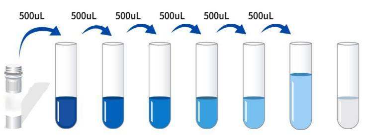 4. Preparation of enzyme conjugate working solution: Before use 15 Minutes will 100× The enzyme conjugate was concentrated in 1000×g centrifugation 1 Minutes, in a general dilution 100× The concentrated enzyme conjugate is diluted to 1× Working concentration ( Example: 10μL Concentrate +990μL Universal diluent ) , ready for use. 5.1× Washing Liquid Preparation: Take 10mL20× Washing liquid to 190mL In distilled water (the concentrated washing liquid taken out of the refrigerator may have crystallization, which is a normal phenomenon. It can be placed at room temperature and prepared after the crystallization is completely dissolved ) 。 V. Operation steps: 1. Equilibration from room temperature 10 After a minute, take out the required slats from the aluminum foil bag, and seal the remaining slats with a ziplock bag and put them back 4℃ 。 2. Sample addition: the samples or standards with different concentrations are separately determined according to 100μL Each well was added to the corresponding well, blank wells were added 100μL Universal diluent. After covering the sealing plate film 37℃ Incubation 60 Minutes. (Recommendation: Minimum dilution of sample to be tested with universal diluent 1 times and then added to the enzyme label plate for testing. Thereby reducing the influence of matrix effect on the test results, and finally multiplying the corresponding dilution factor when calculating the sample concentration. All samples to be tested and standards are recommended to set up double wells in the test). 3. Add biotinylation to detect antibody: take out the enzyme label plate, discard the liquid, and do not wash. Direct addition per well 100μL Biotinochemical test antibody working solution, after covering with sealing film 37℃ Incubation 60 Minutes. 4. Wash the plate: discard the liquid and add each well 300μL 1x Washing liquid, stand 1 Minutes, throw off the washing liquid, pat dry on absorbent paper, and repeat washing the plate 3 Time (plate washer can also be used to wash the plate). 5. Enzyme adding conjugate working solution: each well is added 100μL Enzyme conjugate working solution, covered with sealing membrane 37℃ Incubation 30 Minutes. 6. Wash the plate: discard the liquid according to the steps 4 Washing method, washing plate 5 Twice. 7. Adding substrate: Adding per well 90μL Substrate (TMB) Covered with a sealing film, 37℃ Incubation in the dark 15 Minutes. 8. Adding stop solution: Take out the enzyme label plate and directly add to each well 50μL Stop solution, immediately in 450nm The wavelength of each well was measured OD Value. VI. Calculation of experimental results: Result Judgment: 1. Calculate the average of standard and sample double wells OD Value and subtract the blank well's OD Value as a correction value. Taking the concentration as the abscissa, OD The value is the ordinate , draw a standard curve of a four-parameter logistic function on double logarithmic paper. 2. If the sample OD If the value is above the upper limit of the standard curve, the test should be retested after appropriate dilution and multiplied by the corresponding dilution factor when calculating the sample concentration. 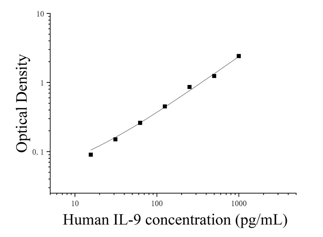 VII. Kit performance: VII. Kit performance: 1. Repeatability: Intraplate coefficient of variation is less than 10% The inter-plate coefficient of variation is less than 10% 。 2. Recovery rate: Add to selected healthy human serum, plasma and cell culture supernatant 3 People with different concentration levels IL-9 , recovery was calculated.
3. Linear dilution: respectively in the selected 4 High concentrations of human serum, plasma and cell culture supernatant were added to healthy human IL-9 Linearity was assessed by dilution within standard curve kinetics.
|
|||||||||||||||||||||||||||||||||||
| Sensitivity | 7.32pg/mL | |||||||||||||||||||||||||||||||||||
| Species Reactivity | Human | |||||||||||||||||||||||||||||||||||
| Theory | This kit uses double antibody sandwich enzyme-linked immunosorbent assay (ELISA). A sample, a standard, a biotin-labeled detection antibody, and a HRP enzyme conjugate were sequentially added to the microwells previously coated with human interleukin 9 (IL-9) capture antibody, incubated and washed in between, and developed color with the substrate TMB, which was converted to blue under the catalysis of peroxidase (HRP) and to final yellow under the action of acid. There was a positive correlation between the shade of color and human interleukin 9 (IL-9) in the sample. The absorbance (OD) was measured by microplate reader at 450nm wavelength, and the concentration of the sample was calculated. | |||||||||||||||||||||||||||||||||||
| Source | Human | |||||||||||||||||||||||||||||||||||
| Synonym | Cytokine P40, HP40, IL9, IL-9, IL-9homolog of mouse T cell and mast cell growth factor 40, interleukin 9, interleukin-9, p40 cytokine, p40 T-cell and mast cell growth factor, P40, T-cell growth factor P40 | |||||||||||||||||||||||||||||||||||
| Detection Type | Double antibody sandwich method | |||||||||||||||||||||||||||||||||||
| Composition |
|
|||||||||||||||||||||||||||||||||||
| Background | Interleukin 9 (IL-9) is a monomeric glycosylated protein with a molecular weight between 28-30 kDa, belonging to the IL-7/IL-9 family, and is a pleiotropic cytokine mainly produced by CD4+ T cells, and mast cells and natural killer cells can also produce IL-9. Initially, IL-9 was thought to be a growth factor of T cells and mast cells. Later, it was found that IL-9 can also regulate the production of B cell immunoglobulins, enhance the expression of mast cell proteases, promote goblet cell proliferation and mucus production, indicating that IL-9 may be involved in the occurrence of allergic inflammatory reactions. IL-9 contains 144 amino acids, including an 18 amino acid signal peptide, and also has 10 cysteine residues and 4 potential N-linked glycosylation sites in the mature IL-9 structure. | |||||||||||||||||||||||||||||||||||
| General Notes | 1. Incubate strictly according to the specified time and temperature to ensure accurate results. All reagents must reach room temperature 20-25°C prior to use. Refrigerate reagents immediately after use. 2. Incorrect washing may lead to inaccurate results. Be sure to absorb as much liquid as possible from the wells before adding the substrate. Do not allow the micropores to dry for too long throughout the process. 3. Clean the residual liquid and fingerprints at the bottom of the board, otherwise it will affect the OD value. 4. The substrate chromogenic solution should be colorless, and the substrate solution that has turned blue cannot be used. 5. Avoid cross-contamination of reagents and samples to avoid wrong results. 6. Avoid direct exposure to strong light during storage and incubation. 7. Any reaction reagent cannot contact the bleaching solvent or the strong gas emitted by the bleaching solvent. Any bleaching ingredient will destroy the biological activity of the reagents in the kit. 8. Expired products cannot be used, and components with different item numbers and batch numbers should not be mixed. 9. Recombinant proteins from sources other than the kit may not match the antibodies of this kit and not be recognized. 10. If it is possible to spread the disease, all samples should be managed well, and samples and detection devices should be processed according to the prescribed procedures. |
|||||||||||||||||||||||||||||||||||
| Storage Temp. | Unopened kit, stored at 2-8℃, valid for 6 months. | |||||||||||||||||||||||||||||||||||
| Test Range | 15.62-1000pg/mL | |||||||||||||||||||||||||||||||||||
| Applications | Cell supernatant, serum, plasma |