1. Isolation of Murine Bone-Marrow Cells
1.1 Execute 6–8-week-old C57BL/6 mice (or strain specified in study plan) by cervical dislocation. Aseptically dissect both femora; remove adhering muscle with sterile scissors/forceps.
1.2 Disinfect bones in 75 % ethanol (2 min), then rinse twice with 10 mL ice-cold PBS.
1.3 Cut off epiphyses; flush marrow cavity with 5 mL cold PBS using a 25 G needle attached to a 10 mL syringe. Repeat until diaphysis appears white.
1.4 Pool marrow suspensions; filter through a 70 µm (200-mesh) nylon cell strainer into a 50 mL tube.
1.5 Centrifuge 1200 rpm (≈ 300 × g), 5 min, 4 °C; discard supernatant.
1.6 Resuspend pellet in 2 mL ACK red-cell lysis buffer; incubate 3–5 min at RT.
1.7 Add 10 mL PBS to stop lysis; centrifuge as above.
1.8 Wash once with 10 mL PBS; resuspend in complete RPMI-1640 + 10 % FBS (pen/strep 1 %, 2 mM L-Gln). Keep on ice.
2. In-vitro Differentiation of BMDC
2.1 Plate 1 × 10⁶ nucleated cells per mL in 6-well plates (3 mL/well) in complete medium supplemented with:
• GM-CSF -20 ng/mL
• IL-4- 20 ng/mL
This is designated Day 0 (D0).
2.2 Semi-media change every 48 h:
a. Remove 1.5 mL supernatant (non-adherent cells) into 15 mL tube.
b. Centrifuge 300 × g, 5 min; resuspend in 1.5 mL fresh complete medium containing fresh cytokines (same concentration).
c. Return cell suspension to original well.
2.3 On D6 harvest loosely adherent and firmly adherent cells:
• Pipette off supernatant; gently rinse wells with 2 mL PBS.
• Add 2 mL cold PBS, scrape with rubber policeman; pool all cells.
• Centrifuge 300 × g, 5 min; count.
• Re-plate at 1 × 10⁶ cells/mL in fresh complete medium + GM-CSF (20 ng/mL) + IL-4 (20 ng/mL).
These cells are “immature BMDC” (iBMDC).
2.4 Maturation (optional): On D8 add:
• TNF-α 20 ng/mL
• GM-CSF & IL-4 as above
Harvest on D10; these are “mature BMDC” (mBMDC).
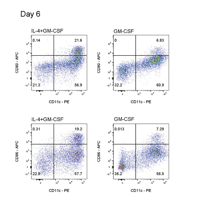
3. Flow-Cytometric Validation of iBMDC (D6)
3.1 Harvest cells as in 2.3; wash once with PBS.
3.2 Count; adjust to 1 × 10⁷ cells/mL in FACS buffer (PBS + 2 % FBS).
3.3 Block Fc receptors: aliquot 100 µL/well (≈ 1 × 10⁶ cells) into 96-well V-bottom plate or FACS tube; add Mouse FcR Blocking Reagent (Mouse IgG); incubate 30 min on ice.
3.4 Stain surface markers (manufacturer’s titration):
Panel 1: PE anti-CD11c (clone N418, Cat. 117307) + APC anti-CD80
Panel 2: PE anti-CD11c (as above) + APC anti-CD86
Incubate 30 min, 4 °C, dark.
3.5 Wash twice with 200 µL FACS buffer; centrifuge 300 × g, 5 min.
3.6 Resuspend in 100 µL FACS buffer; add 7-AAD viability dye 5 µL per tube; incubate 5 min RT, dark.
3.7 Acquire on flow cytometer (≥ 20 000 events per sample).
Expected iBMDC phenotype: CD11c⁺ CD80^low CD86^low 7-AAD⁻.
4. Flow-Cytometric Validation of mBMDC (D10)
4.1–4.7 Identical procedure as Section 3, but use cells harvested after TNF-α maturation.
Expected mBMDC phenotype: CD11c⁺ CD80^high CD86^high 7-AAD⁻.