WB result of HA Tag Recombinant Rabbit mAb
Primary antibody: HA Tag Recombinant Rabbit mAb at 1/10000 dilution
Lane 1: 293T transfected with empty vector whole cell lysate 20 µg
Lane 2: 293T transfected with HA-GFP fusion protein whole cell lysate 20 µg
Secondary antibody: Goat Anti-Rabbit IgG, (H+L), HRP conjugated at 1/10000 dilution
Predicted MW: 28 kDa
Observed MW: 28 kDa
HA Tag Recombinant Rabbit mAb (S-4144)
HA Tag Recombinant Rabbit mAb (S-4144)
Price:
Regular price
$70 USD
Regular price
Sale price
$70 USD
Unit price
per
For shipping services or bulk orders, you may request a quotation.
Secure checkout with

View full details
Product Details
Product Details
Product Specification
| Host | Rabbit |
| Antigen | HA tag |
| Clone Number | S-4144 |
| Antibody Type | Recombinant mAb |
| Isotype | IgG |
| Application | WB, ICC, ICFCM, ChIP |
| Reactivity | Species Independent |
| Purification | Protein A |
| Concentration | 1 mg/ml |
| Conjugation | Unconjugated |
| Physical Appearance | Liquid |
| Storage Buffer | PBS, 40% Glycerol, 0.05% BSA, 0.03% Proclin 300 |
| Stability & Storage | 12 months from date of receipt / reconstitution, -20 °C as supplied |
Dilution
| application | dilution | species |
| WB | 1:5000-1:50000 | Species Independent |
| ICC | 1:1000 | Species Independent |
| ICFCM | 1:1000 | Species Independent |
| ChIP | 1:20-1:50 | Species Independent |
Background
The HA tag (Hemagglutinin tag) is an epitope tag derived from the hemagglutinin (HA) protein of human influenza virus, typically consisting of a 9-amino acid sequence (YPYDVPDYA). Widely used in molecular biology and biochemistry due to its high immunogenicity and specificity, this tag is primarily employed for the detection, purification, and localization of proteins. By genetically fusing the HA tag to the N- or C-terminus of a target protein, high-affinity anti-HA antibodies can be utilized in experiments such as Western blot, co-immunoprecipitation (Co-IP), and immunofluorescence (IF) to effectively track the expression, subcellular localization, and interactions of the target protein. With its small size, high sensitivity, and minimal interference with protein structure, the HA tag has become an essential tool in protein research.
Picture
Picture
Western Blot
FC
Flow cytometric analysis of 4% PFA fixed 90% methanol permeabilized HA tag fusion protein transfected 293F cells (Human embryonic kidney epithelial cell) / (right panel) compared with untransfected 293F cells / (left panel) labeled with anti-HA tag antibody at 1/1000 (0.1 μg) dilution. Then cells were stained with anti-Myc Tag antibody at 1/2000 (0.1 μg) dilution (S0B0209) (Goat Anti - Rabbit IgG Alexa Fluor® 488 was used as the secondary antibody). Flow cytometry and data analysis were performed using Agilent NovoCyte Quanteon and FlowJo™ software.
Immunocytochemistry
ICC shows positive staining in HA tag fusion protein transfected 293F cells (top panel) and negative staining in vector-transfected 293F cells (below panel). Anti- HA Tag antibody was used at 1/1000 dilution (Green) and incubated overnight at 4°C. Goat polyclonal Antibody to Rabbit IgG - H&L (Alexa Fluor® 488) was used as secondary antibody at 1/1000 dilution. The cells were fixed with 100% methanol and permeabilized with 0.1% PBS-Triton X-100. Nuclei were counterstained with DAPI (Blue). Counterstain with tubulin (Red).
ChIP
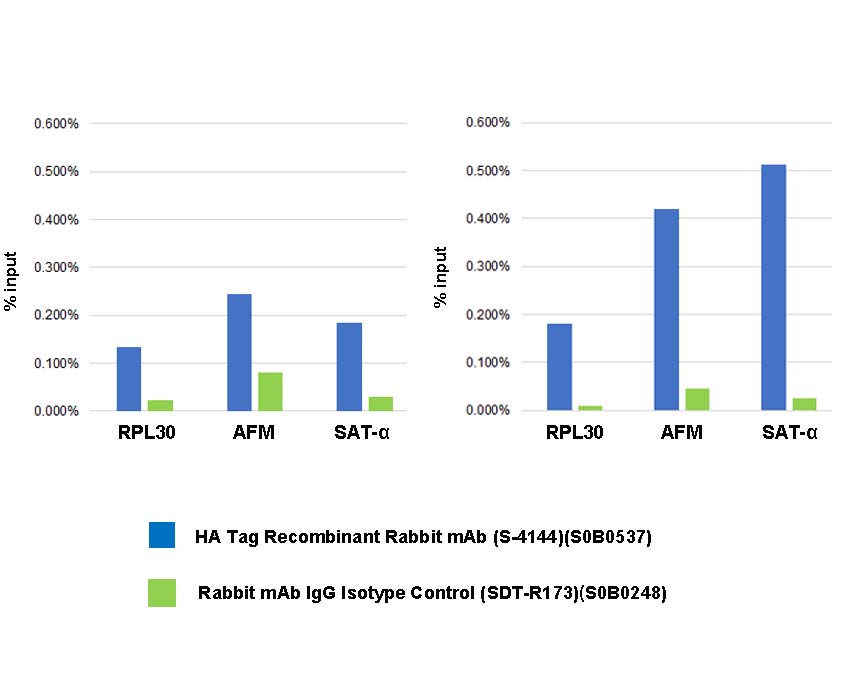
Chromatin immunoprecipitation (ChIP) was performed on 293F cells were either untransfected (left panel) or transfected with an HA-tagged human H3 construct (right panel) cross - linked with 1% formaldehyde for 10 min, then chromatin was fragmented by sonication.
Parallel reactions used HA Tag Recombinant Rabbit mAb (S-4144) and Rabbit mAb IgG Isotype Control (SDT-R173) at 1:50 for immunoprecipitation.
Post - immunoprecipitation, both samples were washed, eluted, and cross - links reversed. Purified DNA was analyzed by qPCR.
qPCR showed the enrichment of RPL30, AFM and SAT-α
in HA Tag Recombinant Rabbit mAb (S-4144)-
immunoprecipitated sample.