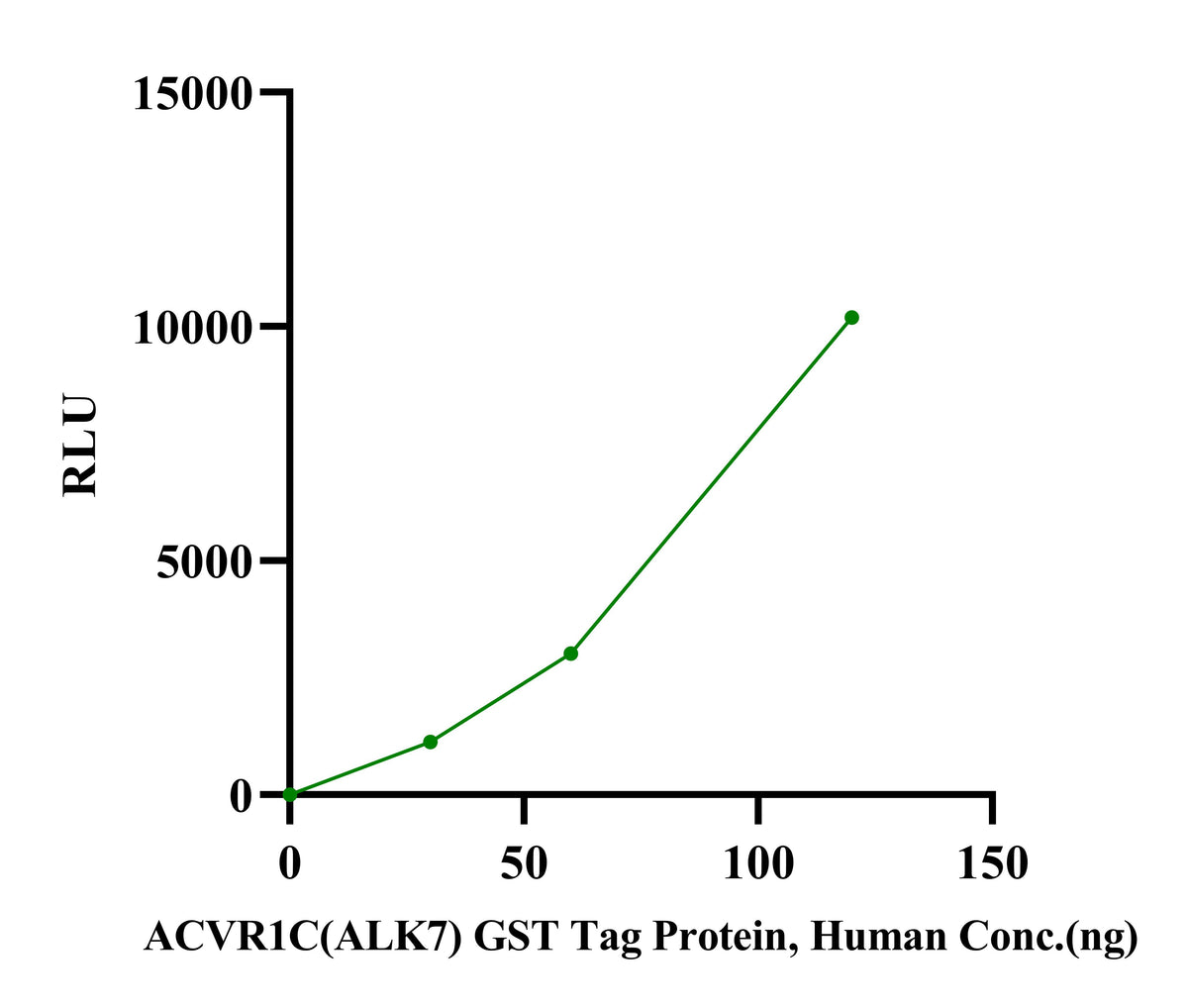
The specific activity of ACVR1C(ALK7) GST Tag Protein, Human was determined to be >3 pmol/min/ug as per activity assay protocol.
ACVR1C(ALK7) GST Tag Protein, Human
ACVR1C(ALK7) GST Tag Protein, Human
Price:
Regular price
$370 USD
Regular price
Sale price
$370 USD
Unit price
per
For shipping services or bulk orders, you may request a quotation.
Secure checkout with

View full details
Product Details
Product Details
Product Specification
| Species | Human |
| Synonyms | ALK-7, ACTR-IC, ACVR1C |
| Accession | Q8NER5 |
| Amino Acid Sequence | Arg139-Ala493 with GST Tag at the N-Terminus |
| Expression System | Baculovirus-InsectCells |
| Molecular Weight | 55-70kDa (Reducing) |
| Purity | >95% by SDS-PAGE |
| Conjugation | Unconjugated |
| Tag | GST Tag |
| Physical Appearance | Liquid |
| Storage Buffer | 50mM Tris, 150mM NaCl, pH7.5, 1mM DTT, 10%Glycerol |
| Stability & Storage | Stable for 12 months upon stored at -80℃ from the date of receipt. And avoid repeated freeze-thaws cycles. |
| Reference | 1. Ibáñez, C.F. (2022). Regulation of metabolic homeostasis by the TGF-β superfamily receptor ALK7. The FEBS Journal, 289(19), 5776-5797. |
Background
ALK7 (Activin Receptor-Like Kinase 7) , officially designated ACVR1C (Activin A Receptor Type 1C) , is a type I receptor serine/threonine kinase belonging to the transforming growth factor-β (TGF-β) superfamily. The gene is located on human chromosome 2q24.1, with the gene symbol ACVR1C and aliases including ALK7 and ACVRLK7.
The full-length ALK7 protein consists of 493 amino acids and exhibits all characteristic features of TGF-β type I receptors, including: conserved cysteines and a putative glycosylation site in the extracellular region, a transmembrane region, a GS box, and a C-terminal serine/threonine-rich kinase domain in the intracellular region. Through alternative splicing, the human ALK7 gene encodes four distinct proteins: full-length ALK7, truncated ALK7 (tALK7), and two soluble forms (sALK7a and sALK7b). The ALK7 gene comprises at least 9 exons and 8 introns.
Protocol
Assay protocol
Principle: The ACVR1C (ALK7) assay is performed using the ADP-GloTM Kinase Assay kit which quantifies the amount of ADP produced by the ACVR1C (ALK7) reaction. The ADP-GloTM Reagent is added to terminate the kinase reaction and to deplete the remaining ATP, and then the Kinase Detection Reagent is added to convert ADP to ATP and to measure the newly synthesized ATP using luciferase/luciferin reaction.
Materials
1.Kinase assay buffer(5X): 200 mM Tris-HCl, pH 7.4, 100 mM MgCl2 and 0.5 mg/mL BSA
2.Kinase Dilution buffer(1X): 40 mM Tris-HCl, pH 7.4, 20 mM MgCl2, 0.1 mg/mL BSA, 1mM DTT
3.ACVR1C (ALK7) GST Tag Protein, Human(Catalog # UA085091)
4.ADP-Glo Kinase Assay (UA, Catalog # UA070101)
5.Substrate: Recombinant Human Smad3 Protein (Full Length, GST Tag) (Sinobiological, Catalog # S12-30G)
6.Solid white multi-well plate (384-well plate) (absin, Catalog # abs7311)
7.Plate Reader (PerkinElmer)
Produce
1.Prepare a substrate/ATP mixture as follows (50 μM example).
Sample Name |
Amount (μL) |
10 mM ATP Solution |
1 |
Kinase Dilution Buffer (1x) |
39 |
Substrate at 200 ng/μL |
40 |
2. Dilute the ACVR1C (ALK7) to 40 µg/mL, 20 µg/mL and 10 µg/mL in Kinase Assay Buffer (1x) and dispense 3 μL into each well of a 384-well plate.
3. Initiate the reaction by adding 2 μL of the detection system prepared in Step 1 to each well. Include a detection system with 3 μL Kinase Assay Buffer (1x) as Blank. The reaction volume is 5 μL.
4. Incubate the reaction at room temperature (22–25℃) for 40 minutes.
5.Add 5 μL of ADP-Glo Reagent to the completed reaction, mix briefly and incubate for 40 minutes at room temperature (22–25 ℃).
6.Add 10 μL of Detection Reagent and incubate the plate for 30 minutes at room temperature (22–25 ℃).
7.Read at luminescence, respectively in endpoint mode.
8.Calculate specific activity.
• Standard Curve
1.Dilute the ATP and ADP to 25 μM in Kinase Assay Buffer (1x).
2.Mix 25 μΜ ATP and 25 μM ADP to form an ATP+ADP solution provided below and dispense 5 μL into each well of a 384-well plate.
Well Number |
1 |
2 |
3 |
4 |
5 |
6 |
7 |
8 |
9 |
10 |
11 |
12 |
25μM ADP (μL) |
100 |
80 |
60 |
40 |
20 |
10 |
5 |
4 |
3 |
2 |
1 |
0 |
25μM ATP (μL) |
0 |
20 |
40 |
60 |
80 |
90 |
95 |
96 |
97 |
98 |
99 |
100 |
3.Add 5 μL of ADP-Glo Reagent to the completed reaction, mix briefly and incubate for 40 minutes at room temperature (22–25 ℃).
4.Add 10 μL of Detection Reagent and incubate the plate for 30 minutes at room temperature (22–25 ℃).
5.Read at luminescence, respectively in endpoint mode.
6.Detect optical signals and establish conversion curves.
Specific Activity (pmol/min/μg) = |
ATP (pmol)-Blank |
Incubation time(min) ×amount of enzyme (μg) |
Picture
Picture
Bioactivity
SDS-PAGE
2μg (R: reducing condition, N: non-reducing condition).