WB result of UBE2I / UBC9 Rabbit pAb
Primary antibody: UBE2I / UBC9 Rabbit pAb at 1/1000 dilution
Lane 1: Jurkat whole cell lysate 20 µg
Lane 2: HeLa whole cell lysate 20 µg
Lane 3: A-172 whole cell lysate 20 µg
Lane 4: Ramos whole cell lysate 20 µg
Secondary antibody: Goat Anti-rabbit IgG (H+L), HRP conjugated at 1/10000 dilution
Predicted MW: 18 kDa
Observed MW: 18 kDa
This blot was developed with high sensitivity substrate
UBE2I / UBC9 Rabbit Polyclonal Antibody
UBE2I / UBC9 Rabbit Polyclonal Antibody
Price:
Regular price
$100 USD
Regular price
Sale price
$100 USD
Unit price
per
For shipping services or bulk orders, you may request a quotation.
Secure checkout with

View full details
Product Details
Product Details
Product Specification
| Host | Rabbit |
| Antigen | UBE2I / UBC9 |
| Synonyms | SUMO-conjugating enzyme UBC9; RING-type E3 SUMO transferase UBC9; SUMO-protein ligase; Ubiquitin carrier protein 9; Ubiquitin carrier protein I; Ubiquitin-conjugating enzyme E2 I; UBCE9 |
| Immunogen | Synthetic Peptide |
| Location | Cytoplasm, Nucleus |
| Accession | P63279 |
| Antibody Type | Polyclonal antibody |
| Isotype | IgG |
| Application | WB, ICC |
| Reactivity | Hu, Ms, Rt, Mk |
| Positive Sample | Jurkat, HeLa, A-172, Ramos, NIH/3T3, mouse spleen, rat spleen, COS-7 |
| Purification | Immunogen Affinity |
| Concentration | 0.5 mg/ml |
| Conjugation | Unconjugated |
| Physical Appearance | Liquid |
| Storage Buffer | PBS, 40% Glycerol, 0.05% BSA, 0.02% sodium azide |
| Stability & Storage | 12 months from date of receipt / reconstitution, -20 °C as supplied |
Dilution
| application | dilution | species |
| WB | 1:1000 | Hu, Ms, Rt, Mk |
| ICC | 1:500 | Hu, Ms, Rt |
Background
UBE2I (also known as UBC9) is the only known SUMO E2 conjugating enzyme in humans, encoded by the UBE2I gene on chromosome 16p13.3. With a molecular weight of approximately 18 kDa, it belongs to the ubiquitin-conjugating E2 enzyme family but specifically recognizes SUMO rather than ubiquitin. In the SUMOylation cascade, the E1 activating enzyme SAE1/SAE2 activates SUMO precursors in an ATP-dependent manner and then transfers mature SUMO1, SUMO2, SUMO3, and other SUMO paralogs to the active cysteine residue Cys93 of UBE2I via a thioester bond. Subsequently, UBE2I recognizes the ΨKxD/E consensus motif on substrate proteins and forms an isopeptide bond between the C-terminal diglycine of SUMO and the ε-amino group of the substrate lysine. This process can be carried out independently by UBE2I or enhanced in specificity and efficiency with the assistance of E3 ligases such as PIAS, RANBP2, CBX4, and ZNF451. UBE2I can also promote poly-SUMO chain formation through non-covalent interactions with SUMO, a process critical for stress adaptation in DNA damage responses and heat shock responses. Functionally, UBE2I-mediated SUMOylation broadly regulates transcription factor activity, chromatin architecture, nuclear pore complex assembly, DNA damage repair, cell cycle progression, and chromosome segregation, serving as a central hub for maintaining nuclear structure and genome stability. Its own activity can be modulated by post-translational modifications such as Ser71 phosphorylation, and its dysfunction is associated with various cancers, neurodegenerative diseases, and immune disorders, making it an important node for targeted therapy of the SUMO pathway.
Picture
Picture
Western Blot
WB result of UBE2I / UBC9 Rabbit pAb
Primary antibody: UBE2I / UBC9 Rabbit pAb at 1/1000 dilution
Lane 1: NIH/3T3 whole cell lysate 20 µg
Secondary antibody: Goat Anti-rabbit IgG (H+L), HRP conjugated at 1/10000 dilution
Predicted MW: 18 kDa
Observed MW: 18 kDa
This blot was developed with high sensitivity substrate
WB result of UBE2I / UBC9 Rabbit pAb
Primary antibody: UBE2I / UBC9 Rabbit pAb at 1/1000 dilution
Lane 1: mouse skeletal muscle lysate 20 µg
Lane 2: mouse spleen lysate 20 µg
Low expression control: mouse skeletal muscle lysate
Secondary antibody: Goat Anti-rabbit IgG (H+L), HRP conjugated at 1/10000 dilution
Predicted MW: 18 kDa
Observed MW: 18 kDa
This blot was developed with high sensitivity substrate
WB result of UBE2I / UBC9 Rabbit pAb
Primary antibody: UBE2I / UBC9 Rabbit pAb at 1/1000 dilution
Lane 1: PC-12 whole cell lysate 20 µg
Lane 2: rat spleen lysate 20 µg
Secondary antibody: Goat Anti-rabbit IgG (H+L), HRP conjugated at 1/10000 dilution
Predicted MW: 18 kDa
Observed MW: 18 kDa
This blot was developed with high sensitivity substrate
WB result of UBE2I / UBC9 Rabbit pAb
Primary antibody: UBE2I / UBC9 Rabbit pAb at 1/1000 dilution
Lane 1: COS-7 whole cell lysate 20 µg
Secondary antibody: Goat Anti-rabbit IgG (H+L), HRP conjugated at 1/10000 dilution
Predicted MW: 18 kDa
Observed MW: 18 kDa
This blot was developed with high sensitivity substrate
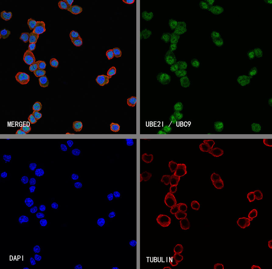
Immunocytochemistry
ICC shows positive staining in Jurkat cells. Anti-UBE2I / UBC9 antibody was used at 1/500 dilution (Green) and incubated overnight at 4°C. Goat polyclonal Antibody to Rabbit IgG - H&L (Alexa Fluor® 488) was used as secondary antibody at 1/1000 dilution. The cells were fixed with 4% PFA and permeabilized with 0.1% PBS-Triton X-100. Nuclei were counterstained with DAPI (Blue). Counterstain with tubulin (Red).
ICC shows positive staining in NIH/3T3 cells. Anti-UBE2I / UBC9 antibody was used at 1/500 dilution (Green) and incubated overnight at 4°C. Goat polyclonal Antibody to Rabbit IgG - H&L (Alexa Fluor® 488) was used as secondary antibody at 1/1000 dilution. The cells were fixed with 4% PFA and permeabilized with 0.1% PBS-Triton X-100. Nuclei were counterstained with DAPI (Blue). Counterstain with tubulin (Red).
ICC shows positive staining in PC-12 cells. Anti-UBE2I / UBC9 antibody was used at 1/500 dilution (Green) and incubated overnight at 4°C. Goat polyclonal Antibody to Rabbit IgG - H&L (Alexa Fluor® 488) was used as secondary antibody at 1/1000 dilution. The cells were fixed with 4% PFA and permeabilized with 0.1% PBS-Triton X-100. Nuclei were counterstained with DAPI (Blue). Counterstain with tubulin (Red).