WB result of Neutrophil Elastase Recombinant Rabbit mAb
Primary antibody: Neutrophil Elastase Recombinant Rabbit mAb at 1/1000 dilution
Lane 1: mouse bone marrow lysate 20 µg
Secondary antibody: Goat Anti-rabbit IgG (H+L), HRP conjugated at 1/10000 dilution
Predicted MW: 28 kDa
Observed MW: 30 kDa
Neutrophil Elastase Recombinant Rabbit mAb (S-4397-22)
Neutrophil Elastase Recombinant Rabbit mAb (S-4397-22)
Price:
Regular price
$100 USD
Regular price
Sale price
$100 USD
Unit price
per
For shipping services or bulk orders, you may request a quotation.
Secure checkout with

View full details
Product Details
Product Details
Product Specification
| Host | Rabbit |
| Antigen | Neutrophil Elastase |
| Synonyms | Neutrophil elastase; Elastase-2; Leukocyte elastase; Ela2; Elane |
| Immunogen | Synthetic Peptide |
| Location | Nucleus, Lysosome |
| Accession | Q3UP87 |
| Clone Number | S-4397-22 |
| Antibody Type | Recombinant mAb |
| Isotype | IgG |
| Application | WB, IHC-P, IF |
| Reactivity | Ms |
| Purification | Protein A |
| Concentration | 0.5 mg/ml |
| Conjugation | Unconjugated |
| Physical Appearance | Liquid |
| Storage Buffer | PBS, 40% Glycerol, 0.05% BSA, 0.02% sodium azide |
| Stability & Storage | 12 months from date of receipt / reconstitution, -20 °C as supplied |
Dilution
| application | dilution | species |
| WB | 1:1000 | Ms |
| IHC-P | 1:1000 | Ms |
| ICC | 1:500 | Ms |
Background
Neutrophil elastase (NE; gene ELANE, also known as ELA2) is a 218-amino-acid, ~29–33 kDa chymotrypsin-like serine protease that is synthesized in promyelocytes and stored in the azurophilic (primary) granules of neutrophil granulocytes. It is released upon neutrophil activation and degrades elastin as well as collagen, fibronectin, proteoglycans, and other matrix and plasma proteins, giving it a central role in antimicrobial defense, inflammatory tissue injury, and matrix remodeling. Its proteolytic activity is tightly controlled by endogenous inhibitors, chiefly α1-antitrypsin (SERPINA1), and inherited ELANE mutations cause severe congenital neutropenia, while unchecked elastase activity contributes to emphysema, ARDS, and chronic inflammatory lung disease. In diagnostic pathology, NE is used as a sensitive immunohistochemical marker of neutrophil/myeloid lineage (e.g., clone NP57), helpful in identifying neutrophilic infiltrates, myeloid leukemia, and neutrophil-rich skin or soft-tissue lesions.
Picture
Picture
Western Blot
Immunohistochemistry
IHC shows positive staining in paraffin-embedded mouse bone marrow. Anti-Neutrophil Elastase antibody was used at 1/1000 dilution, followed by a HRP Polymer for Rabbit IgG (ready to use). Counterstained with hematoxylin. Heat mediated antigen retrieval with Tris/EDTA buffer pH9.0 was performed before commencing with IHC staining protocol.
IHC shows positive staining in paraffin-embedded mouse spleen. Anti-Neutrophil Elastase antibody was used at 1/1000 dilution, followed by a HRP Polymer for Rabbit IgG (ready to use). Counterstained with hematoxylin. Heat mediated antigen retrieval with Tris/EDTA buffer pH9.0 was performed before commencing with IHC staining protocol.
Negative control: IHC shows negative staining in paraffin-embedded mouse brain. Anti- Neutrophil Elastase antibody was used at 1/1000 dilution, followed by a HRP Polymer for Rabbit IgG (ready to use). Counterstained with hematoxylin. Heat mediated antigen retrieval with Tris/EDTA buffer pH9.0 was performed before commencing with IHC staining protocol.
Immunofluorescence
IF shows positive staining in paraffin-embedded mouse bone marrow. Anti- Neutrophil Elastase antibody was used at 1/500 dilution (Green) and incubated overnight at 4°C. Goat polyclonal Antibody to Rabbit IgG - H&L (Alexa Fluor® 488) was used as secondary antibody at 1/1000 dilution. Counterstained with DAPI (Blue). Heat mediated antigen retrieval with Tris/EDTA buffer pH9.0 was performed before commencing with IF staining protocol.
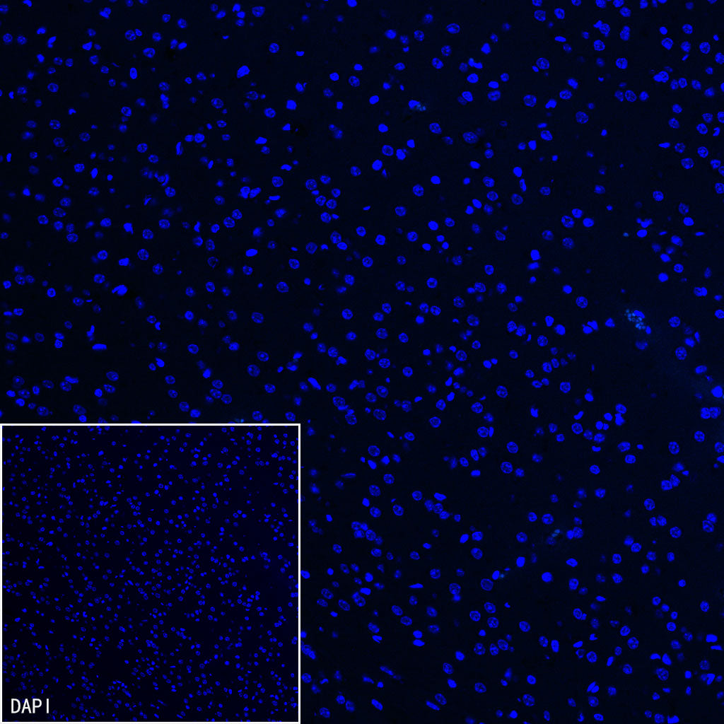
Negative control: IF shows negative staining in paraffin-embedded mouse brain. Anti- Neutrophil Elastase antibody was used at 1/500 dilution and incubated overnight at 4°C. Goat polyclonal Antibody to Rabbit IgG - H&L (Alexa Fluor® 488) was used as secondary antibody at 1/1000 dilution. Counterstained with DAPI (Blue). Heat mediated antigen retrieval with Tris/EDTA buffer pH9.0 was performed before commencing with IF staining protocol.