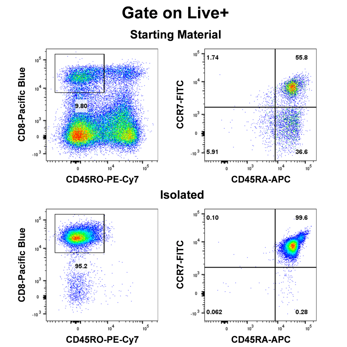
Human naïve CD8 T cells were enriched from peripheral blood mononuclear cells (PBMCs). After sorting, the cells were stained with the following fluorescent antibodies: CD8-Pacific Blue (Clone: S-R533), CD45RO-PE-Cy7 (Clone: UCHL1), CD45RA-APC (Clone: HI100), and CCR7 (CD197)-FITC (Clone: S-R704). Gating was performed on live cells (Live⁺) for analysis, and the proportion of naïve CD8 T cells in the sorted fraction was determined. Flow cytometry results showed that the purity of CD8⁺CD45RO⁻ cells after sorting was 95.2%, and the purity of CD45RA⁺CCR7⁺ cells within the CD8⁺CD45RO⁻ population was 99.6%.
Human Naïve CD8 T Cell Isolation Kit (Column-Based)
Human Naïve CD8 T Cell Isolation Kit (Column-Based)
Price:
Regular price
$1,085 USD
Regular price
Sale price
$1,085 USD
Unit price
per
For shipping services or bulk orders, you may request a quotation.
Secure checkout with

View full details
Product Details
Product Details
Product Specification
| Format |
1 mL Human Naïve CD8 T Cell Biotin-Antibody Cocktail; 1.5 mL Streptavidin Beads; All components are stored in a buffer containing 0.05% sodium azide. |
| Capacity | Suitable for a total cell count of 1 × 10⁹, and can perform up to 100 sorting runs. |
| Principle of Separation | This kit employs a column-based negative selection method to isolate Human Naïve CD8 T cells from human peripheral blood mononuclear cells (PBMCs). In this negative selection process, non-Human Naïve CD8 T cells are labeled with biotin-conjugated antibodies, and the cells are then incubated with streptavidin-nanomagnetic beads. As a result, the unwanted cells become bound to the magnetic beads via the antibody complex, while the target Human Naïve CD8 T cells remain unlabeled. The cell suspension is subsequently loaded onto a separation column; under the influence of a magnetic field, the magnetically labeled non-target cells are retained in the column, whereas the unlabeled Human Naïve CD8 T cells elute by gravity into the collection tube. The resulting supernatant represents the enriched fraction of Human Naïve CD8 T cells after removal of the non-target cells. |
| Applications in cell sorting | Naïve CD8 T cells in their unmanipulated, native state are isolated from human PBMCs and can be used for a variety of downstream functional assays. |
| Reactivity | Human |
| Magnetic Bead Size | 20 nm |
| Separation Method | Column-Based |
| Selection Strategy | Negative Selection |
| Stability & Storage | Store protected from light at 2–8°C; do not freeze. |
Background
Human naive CD8⁺ T cells are the core initiating cell population of the adaptive immune response, referring to a subset of CD8⁺ T cells that have not encountered specific antigens and remain in a quiescent state, typically characterized by the phenotype CD8⁺CD45RO⁻CD45RA⁺CCR7⁺. Upon antigen stimulation and cytokine regulation, these cells can be activated and proliferate, differentiating into effector and memory CD8⁺ T cells. They play a crucial role in research on anti-infectious immunity, tumor immunity, and autoimmune diseases, making them an important subject of study in the field of adaptive immunity. The Human Naive CD8⁺ T Cell Negative Selection Kit is developed based on immunomagnetic bead–mediated negative selection technology, which avoids the antibody-binding labeling defects inherent in positive selection. The antibody components in the separation system precisely target surface markers on non-naive CD8⁺ T cells, specifically depleting non-target cell populations such as CD4⁺ T cells, memory CD8⁺ T cells, B cells, NK cells, monocytes, granulocytes, and erythrocytes. The resulting high-purity human naive CD8⁺ T cells are free from exogenous labels and magnetic bead residues, maintaining their native state and preserving their biological potential for proliferation and differentiation to the greatest extent. They are fully compatible with various in vitro functional assays and represent the mainstream tool for isolating and purifying human naive CD8⁺ T cells in current research.
Protocol
Note: This kit does not include the separation buffer; it must be provided separately. The corresponding Stater product number isS0D3018.
Taking the L Separation Column as an example
Steps |
Operating instructions |
Dosage and time |
|
Cell processing and labeling |
1 |
Isolate PBMCs from peripheral blood by density gradient centrifugation using lymphocyte separation medium, or directly thaw cryopreserved PBMCs, and perform cell counting; Note:It is recommended to count cells after staining with AO/PI; PBMCs can be centrifuged at 400 g for 7 minutes; |
Sample preparation |
2 |
Resuspend 1×10⁷cells in 100 μL of MagSep Separation Buffer; Note:Adjust reagent volumes proportionally according to sample volume; retain a pre-sorting sample to determine the proportion of human naïve CD8 T cells; |
1×10⁷cells/100 μL |
|
3 |
Add 10 μL of the Human Naïve CD8 T Cell Biotin-Antibody Cocktail to the sample and gently mix. Note:For this step, gently pipette up and down 2-3 times to mix. |
10 μL/100 μL |
|
4 |
Incubate the antibodies in the cocktail with the cells at 4°C or room temperature for 10 minutes. |
Incubate at 4°C or room temperature for 10 minutes. |
|
5 |
After antibody incubation, add twice the volume of MagSep Separation Buffer, centrifuge at 400 g for 7 minutes, then carefully discard the supernatant. Resuspend 1×10⁷ cells in 100 μL of MagSep Separation Buffer for subsequent steps.Add twice the volume of buffer to wash away the antibodies. |
Add twice the volume of buffer to wash the antibodies. |
|
6 |
Mix well.Streptavidin Beads, add 15 μLof Streptavidin Beadsto the sample; |
15 μL/100 μL |
|
7 |
Gently mix the magnetic beads and cells, and incubate at 4°C or room temperature for 8 minutes; Note:In this step, gently pipette up and down 2-3 times to mix; During the incubation of the magnetic beads, if cells settle, you can mix again after 4 minutes; |
Incubate at 4°C or room temperature for 8 minutes |
|
Cell sorting |
8 |
Place the L Separation Column into the column separation magnet.Add 3 mL of MagSep Separation Buffer to rinse the separation column;Rinse the separation column |
Rinse the separation column |
9 |
Add the cells to the separation column and collect the target cells using a collection tube; Note:The cell volume should be at least 500 μL; if less, add MagSep Separation Buffer to make up the volume. The cells that drip down by gravity are the target cells; |
Collect the target cells |
|
10 |
Add 3 mL of MagSep Separation Buffer to collect the target cells; Note:This step elutes the unmarked target cells from the separation column, increasing the yield. |
Collect the target cells |
|
|
Notes:1. Pay attention to the maximum capacity of the separation column.The maximum number of cells that can be labeled in an S Separation Column is1×10⁷;the maximum number of cells that can be labeled in an L Separation Column is1×10⁸; 2. If the viability of the sample is low, it is recommended to use a dead cell removal kit to eliminate dead cells (Dead Cell Removal Set, (RUO)#S0K0009). | |||
Picture
Picture
Validation Data