WB result of Annexin-4/ANXA4 Recombinant Rabbit mAb
Primary antibody: Annexin-4/ANXA4 Recombinant Rabbit mAb at 1/1000 dilution
Lane 1: HeLa whole cell lysate 20 μg
Lane 2: Hep G2 whole cell lysate 20 μg
Secondary antibody: Goat Anti-Rabbit IgG (H+L), HRP conjugated at 1/10000 dilution
Predicted MW: 36 kDa
Observed MW: 31 kDa
Annexin-4/ANXA4 Recombinant Rabbit mAb (S-6264)
Annexin-4/ANXA4 Recombinant Rabbit mAb (S-6264)
Price:
Regular price
$100 USD
Regular price
Sale price
$100 USD
Unit price
per
For shipping services or bulk orders, you may request a quotation.
Secure checkout with

View full details
Product Details
Product Details
Product Specification
| Host | Rabbit |
| Antigen | Annexin-4/ANXA4 |
| Synonyms | Annexin A4; 35-beta calcimedin; Annexin IV; Carbohydrate-binding protein p33/p41; Chromobindin-4; ANX4 |
| Location | Cytoplasm |
| Accession | P09525 |
| Clone Number | S-6264 |
| Antibody Type | Recombinant mAb |
| Isotype | IgG |
| Application | WB, IHC-P, ICC |
| Reactivity | Hu, Ms, Rt |
| Positive Sample | HeLa, Hep G2, RAW264.7, NIH/3T3, mouse liver, mouse kidney, mouse lung, C6, PC-12, rat liver, rat kidney, rat lung |
| Purification | Protein A |
| Concentration | 0.25 mg/ml |
| Conjugation | Unconjugated |
| Physical Appearance | Liquid |
| Storage Buffer | PBS, 40% Glycerol, 0.05% BSA, 0.02% sodium azide |
| Stability & Storage | 12 months from date of receipt / reconstitution, -20 °C as supplied |
Dilution
| application | dilution | species |
| WB | 1:1000-1:2000 | Hu, Ms, Rt |
| IHC-P | 1:2500 | Hu, Ms, Rt |
| ICC | 1:250 | Hu |
Background
Annexin A4 (ANXA4), also known as lipocortin IV or 36 kDa zymogen granule membrane-associated protein (ZAP36), is a calcium- and phospholipid-binding protein belonging to the annexin superfamily, encoded by the ANXA4 gene. Structurally, it contains a conserved C-terminal core domain composed of four homologous annexin repeats that mediate calcium-dependent phospholipid binding, and a short variable N-terminal domain that is subject to post-translational modifications such as phosphorylation and is thought to confer functional specificity. Annexin A4 is predominantly expressed in epithelial cells and is also found in secretory tissues including the pancreas, liver, intestine, and kidney. Functionally, it promotes calcium-dependent membrane aggregation and fusion, participating in exocytosis and membrane repair, and its lipid association is modulated by sodium ions, which induce dissociation from membranes. Beyond its physiological roles, Annexin A4 is overexpressed in numerous malignancies—including renal cell carcinoma, malignant mesothelioma, endometrial carcinoma, and cholangiocarcinoma—where it contributes to tumor progression and chemoresistance, particularly to cisplatin; notably, its expression is increased in cisplatin-resistant mesothelioma cells, and knockdown of ANXA4 restores drug sensitivity, supporting its potential as a biomarker and therapeutic target.
Picture
Picture
Western Blot
WB result of Annexin-4/ANXA4 Recombinant Rabbit mAb
Primary antibody: Annexin-4/ANXA4 Recombinant Rabbit mAb at 1/1000 dilution
Lane 1: Raw 264.7 whole cell lysate 20 μg
Lane 2: NIH/3T3 whole cell lysate 20 μg
Lane 3: mouse liver lysate 20 μg
Lane 4: mouse kidney lysate 20 μg
Lane 5: mouse lung lysate 20 μg
Secondary antibody: Goat Anti-Rabbit IgG (H+L), HRP conjugated at 1/10000 dilution
Predicted MW: 36 kDa
Observed MW: 31 kDa
WB result of Annexin-4/ANXA4 Recombinant Rabbit mAb
Primary antibody: Annexin-4/ANXA4 Recombinant Rabbit mAb at 1/1000 dilution
Lane 1: C6 whole cell lysate 20 μg
Lane 2: PC-12 whole cell lysate 20 μg
Lane 3: rat liver lysate 20 μg
Lane 4: rat kidney lysate 20 μg
Lane 5: rat lung lysate 20 μg
Secondary antibody: Goat Anti-Rabbit IgG (H+L), HRP conjugated at 1/10000 dilution
Predicted MW: 36 kDa
Observed MW: 31 kDa
Immunohistochemistry
IHC shows positive staining in paraffin-embedded human kidney. Anti-Annexin-4/ANXA4 antibody was used at 1/2500 dilution, followed by a HRP Polymer for Rabbit IgG (ready to use). Counterstained with hematoxylin. Heat mediated antigen retrieval with Tris/EDTA buffer pH9.0 was performed before commencing with IHC staining protocol.
IHC shows positive staining in paraffin-embedded human pancreas. Anti-Annexin-4/ANXA4 antibody was used at 1/2500 dilution, followed by a HRP Polymer for Rabbit IgG (ready to use). Counterstained with hematoxylin. Heat mediated antigen retrieval with Tris/EDTA buffer pH9.0 was performed before commencing with IHC staining protocol.
IHC shows positive staining in paraffin-embedded human prostate. Anti-Annexin-4/ANXA4 antibody was used at 1/2500 dilution, followed by a HRP Polymer for Rabbit IgG (ready to use). Counterstained with hematoxylin. Heat mediated antigen retrieval with Tris/EDTA buffer pH9.0 was performed before commencing with IHC staining protocol.
IHC shows positive staining in paraffin-embedded human cervical squmouas cell carcinoma. Anti-Annexin-4/ANXA4 antibody was used at 1/2500 dilution, followed by a HRP Polymer for Rabbit IgG (ready to use). Counterstained with hematoxylin. Heat mediated antigen retrieval with Tris/EDTA buffer pH9.0 was performed before commencing with IHC staining protocol.
IHC shows positive staining in paraffin-embedded human lung cancer. Anti-Annexin-4/ANXA4 antibody was used at 1/2500 dilution, followed by a HRP Polymer for Rabbit IgG (ready to use). Counterstained with hematoxylin. Heat mediated antigen retrieval with Tris/EDTA buffer pH9.0 was performed before commencing with IHC staining protocol.
IHC shows positive staining in paraffin-embedded mouse stomach. Anti-Annexin-4/ANXA4 antibody was used at 1/2500 dilution, followed by a HRP Polymer for Rabbit IgG (ready to use). Counterstained with hematoxylin. Heat mediated antigen retrieval with Tris/EDTA buffer pH9.0 was performed before commencing with IHC staining protocol.
IHC shows positive staining in paraffin-embedded rat stomach. Anti-Annexin-4/ANXA4 antibody was used at 1/2500 dilution, followed by a HRP Polymer for Rabbit IgG (ready to use). Counterstained with hematoxylin. Heat mediated antigen retrieval with Tris/EDTA buffer pH9.0 was performed before commencing with IHC staining protocol.
Immunocytochemistry
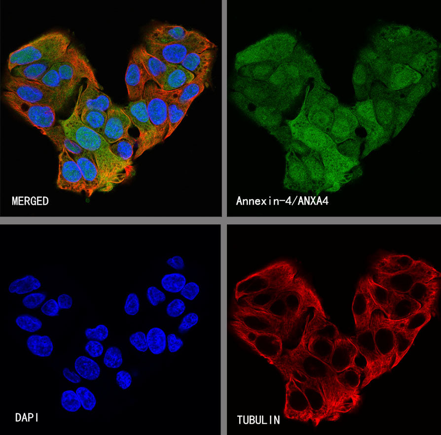
ICC shows positive staining in Hep G2 cells. Anti- Annexin-4/ANXA4 antibody was used at 1/250 dilution (Green) and incubated overnight at 4°C. Goat polyclonal Antibody to Rabbit IgG - H&L (Alexa Fluor® 488) was used as secondary antibody at 1/1000 dilution. The cells were fixed with 4% PFA and permeabilized with 0.1% PBS-Triton X-100. Nuclei were counterstained with DAPI (Blue). Counterstain with tubulin (Red).