
Basement‑Membrane Matrix: Versatile Tools for Organoid Culture, Angiogenesis Assays, In‑Vivo Tumorigenesis and Stem‑Cell Research

Modern life‑science research has moved well beyond conventional two‑dimensional cell culture. Patient‑derived tumor organoids (PDOs), patient‑derived xenografts (PDX), angiogenesis, stem‑cell differentiation, and tumor invasion‑metastasis represent heavily investigated hot topics featured in top‑tier journals. Basement‑membrane matrix serves as a core substrate for nearly all in‑vitro and in‑vivo models recapitulating physiological microenvironments, as reported in Nature, Cell family journals, Cancer Research, Cell Discovery and other high‑impact publications.
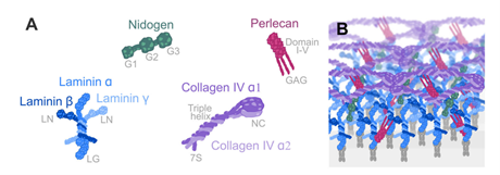
Figure 1. Protein composition and structure of native basement membrane
Nevertheless, researchers frequently encounter technical bottlenecks during reagent selection: endogenous growth‑factor interference in organoid assays, matrix collapse during in‑vivo tumor‑formation studies, high phenol‑red background for fluorescence imaging, and batch‑to‑batch instability in stem‑cell culture. AntBio delivers a comprehensive portfolio of basement‑membrane matrix products, including standard‑formulation, low‑growth‑factor, high‑concentration, iPSC‑validated, and ready‑to‑use variants. These reagents fulfill experimental requirements ranging from basic cell assays to high‑impact translational research.
1. Basement‑Membrane Matrix: A Biologically Active Scaffold for Biological Assays
Basement‑membrane matrix (Matrigel) is a solubilized basement‑membrane extract derived from mouse tumors. Upon incubation at 37 °C, this preparation polymerizes into a biologically‑functional three‑dimensional scaffold that recapitulates the structural, compositional, physical and functional properties of native cellular basement membranes.
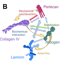
Figure 2. Protein‑protein interactions within basement‑membrane components
Though biochemically complex, its major constituents are well‑defined: ~60 % laminin, ~30 % type‑IV collagen, and ~8 % nidogen. Laminin and type‑IV collagen provide mechanical structural support, while nidogen acts as a bridging molecule to organize extracellular‑matrix assemblies. Additional components include heparan‑sulfate proteoglycans and multiple growth factors: VEGF, TGF‑β, EGF, IGF and FGF.
Key property: thermoreversible gelation. The material remains transparent and liquid at 4 °C, and undergoes spontaneous cross‑linking to form stable 3D hydrogels after 15‑60 min incubation at 37 °C. Gels revert to liquid upon cooling back to 4 °C, facilitating cell recovery and organoid passaging, and supporting diverse 3D in‑vitro and in‑vivo modelling workflows.
2. Core Applications: From 2D to 3D, In‑Vitro to In‑Vivo
2.1 3D Organoid Culture
Organoids are self‑assembled three‑dimensional structures generated from stem or progenitor cells, recapitulating key architectural and functional features of corresponding native organs. Basement‑membrane matrix acts as the indispensable 3D scaffold for generating intestinal, hepatic, gastric, pulmonary and tumor‑derived organoids.
Key experimental workflow
- Digest tissue samples to obtain cell aggregates; recommended volume ratio of matrix gel to cell pellet = 25:1.
- Perform spotting on ice: apply 25‑30 μL matrix droplets per well in 24‑well plates, then incubate at 37 °C for 15‑30 min for gel polymerization.
- Overlay with organoid‑specific medium for long‑term cultivation, passaging and high‑throughput drug screening.
Technical challenges & product selection
Organoids are highly sensitive to intrinsic growth‑factor background; excess endogenous growth factors cause aberrant proliferation and perturbed differentiation. Phenol‑red‑free formulations are required for fluorescence imaging.
✅ Recommendation: abs9495, Low‑Growth‑Factor, Phenol‑Red‑Free, cited in 16 publications.

Figure 4. Side‑by‑side comparison of colorectal‑cancer organoids cultured with different commercial matrix products. AntBio low‑growth‑factor matrix yields larger and more numerous viable colorectal‑cancer organoids compared with a leading brand.
2.2 In‑Vitro Angiogenesis Assays
The in‑vitro angiogenesis assay is a canonical platform for evaluating pro‑ or anti‑angiogenic drug candidates. Human umbilical vein endothelial cells (HUVEC) seeded atop matrix gels rapidly assemble into capillary‑like networks within hours. Quantification of total tube length, branching‑point density and lumen area enables efficient screening of angiomodulatory compounds.
Key workflow: Starve HUVEC for 24 h prior to seeding onto matrix thin layers; assess capillary‑network formation within 6‑12 h for angiogenesis research and anti‑angiogenic drug screening.
✅ Recommendation: Standard‑formulation abs9490 / abs9491. Uniform tube formation, intact vascular networks and high imaging quality; cited in 18 publications.

Figure 5. HUVEC capillary‑network formation on AntBio OrganoGel and competitor matrix. Live‑cell staining performed with Calcein‑AM (green). Scale bar = 100 μm.
2.3 Subcutaneous Tumorigenesis in Mice
This assay implants human‑derived tumor cells into immunodeficient mice to monitor tumour initiation and progression. Immunodeficient strains are required to mitigate immune rejection. Tumour cells are delivered subcutaneously (easy manipulation and monitoring) or orthotopically (higher physiological relevance). Tumour‑volume measurements enable therapeutic‑efficacy assessment and mechanistic tumour‑biology studies.
Key workflow
- Mix logarithmically‑growing tumour‑cell suspensions with matrix gel at a 1:1 ratio on ice (4 °C).
- Maintain low‑temperature conditions throughout the procedure to prevent premature gelation.
- Inject the cell‑matrix mixture subcutaneously (commonly axillary region) into 4‑6‑week‑old mice.
- Monitor tumour formation; palpable tumours typically emerge within 1‑2 weeks.
Important Notes
- Animal husbandry: Use 4‑6‑week‑old Nude mice; acclimate animals under SPF housing for one week before experiments.
- Injection sites and cell dosage: Common routes include subcutaneous, intravenous and orthotopic administration. Typical cell dosage ranges from 1‑5 × 10⁶ cells per 200 μL; adjust according to individual cell‑line characteristics.
- Tumour‑onset timeline: Subcutaneous tumours generally develop within 1‑2 weeks (maximum 1 month). Intravenous or intraperitoneal implants usually form tumours within ~1 week. Closely monitor body weight and general health status.
- Avoid leakage or intramuscular injection, which causes poor tumour‑volume consistency. Advance the needle tip upward during injection; keep injection volume ≤ 200 μL.
✅ Recommendation: abs9493, High‑Concentration, Phenol‑Red‑Free, cited in 9 publications.
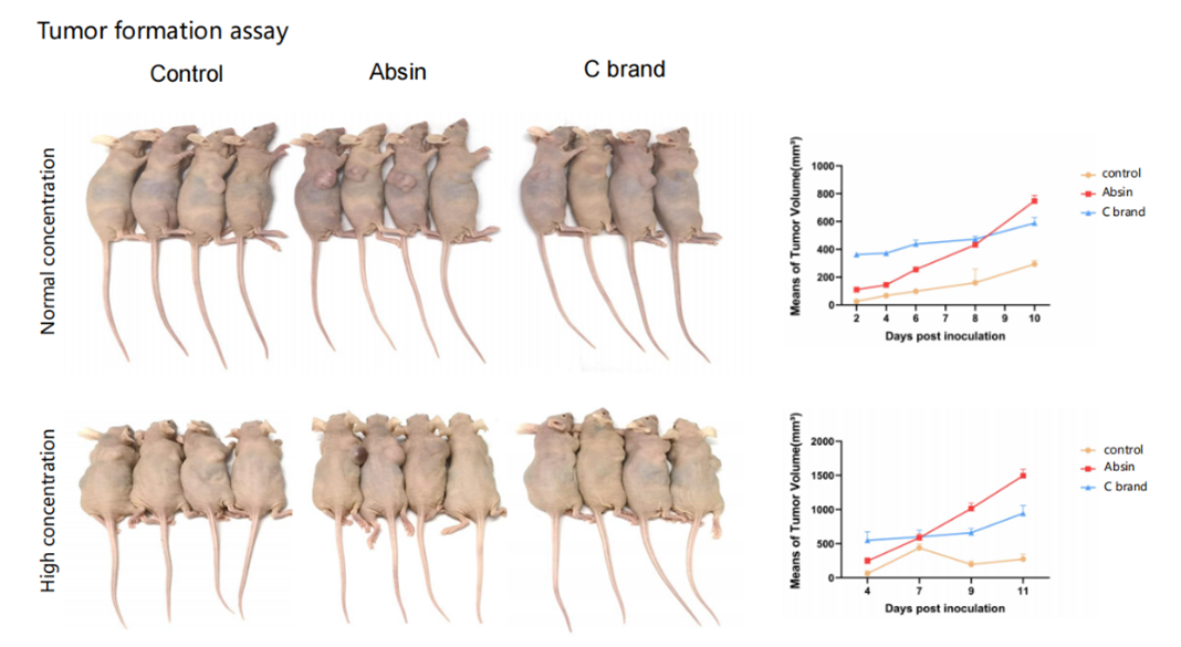
Figure 6. In‑vivo tumour‑formation assay. Left: representative nude‑mouse images; right: quantitative tumour‑volume growth curves. Both normal‑ and high‑concentration AntBio matrix groups achieve larger late‑stage subcutaneous tumour volumes than control and brand‑C groups.
2.4 Culture and Differentiation of Human Pluripotent Stem‑Cells
Basement‑membrane matrix coating represents the gold‑standard feeder‑free culture system for maintaining pluripotency of human embryonic stem cells (hESCs) and induced pluripotent stem cells (iPSCs).
Key workflow
- Thaw matrix overnight in a 4 °C ice bath; gently pipette 5 times with pre‑chilled pipette tips to homogenize. Minimize bubble formation; remove residual bubbles by brief low‑speed centrifugation if needed.
- Pre‑warm culture plates inside a cell incubator.
- Aliquot thawed matrix using pre‑chilled pipette tips.
- Dilute stock matrix at 1:100 with ice‑cold serum‑free medium, spread evenly across plate surfaces. Recommended coating volume: 300 μL/cm².
- Incubate coated plates at room temperature for 1 h.
- Remove coating solution and immediately seed stem cells suspended in mTeSR1 medium. Prevent complete drying of coated surfaces.
✅ Recommendation: abs9496, iPSC‑Validated, Phenol‑Red‑Free.

Figure 7. Functional scoring comparison across multiple matrix suppliers. abs9496 exhibits performance comparable to leading commercial products.
2.5 High‑Throughput Coating
Conventional basement‑membrane matrix requires 4 °C aliquoting and ice‑cold handling, which frequently causes premature gelation and reagent waste for novice users, alongside long processing times for high‑throughput screening and large‑scale coating. AntBio abs9410 Ready‑to‑Use Matrix eliminates freezing‑thawing and dilution steps for streamlined workflows and improved experimental throughput.
Key workflow (6‑well plate example): Add 1‑2 mL ready‑to‑use matrix per well and rock plates to fully cover well bottoms. Incubate plates for 1‑2 h at 37 °C under 5 % CO₂. For delayed usage, seal plates with Parafilm and store at 2‑8 °C for up to one week.
Prior to cell seeding, equilibrate pre‑coated plates at room temperature inside biosafety cabinets for 20 min. Aspirate excess matrix, then seed cells promptly.
3. AntBio Basement‑Membrane Matrix Portfolio
| Catalog No. | Product Name | Specification | Applications |
|---|---|---|---|
| abs9490 | Standard OrganoGel with Phenol red | 1.5 mL × 4 / 1.5 mL × 8 | Angiogenesis assays; cell‑invasion assays |
| abs9491 | Standard OrganoGel Phenol red free | 1.5 mL × 8 | Angiogenesis assays; cell‑invasion assays |
| abs9492 | HC OrganoGel with Phenol red | 1.5 mL × 8 | Mouse subcutaneous tumorigenesis; gel‑plug embolism assays |
| abs9493 |
HC OrganoGel Phenol red free |
1.5 mL × 4 / 1.5 mL × 8 | Mouse subcutaneous tumorigenesis; gel‑plug embolism assays |
| abs9494 | GFR OrganoGel with Phenol red | 1.5 mL × 4 / 1.5 mL × 8 | Organoid culture; growth‑factor and signalling‑pathway studies |
| abs9495 | GFR OrganoGel Phenol red free | 1.5 mL × 4 / 1.5 mL × 8 | Organoid culture; growth‑factor and signalling‑pathway studies |
| abs9497 | HC&GFR OrganoGel with Phenol red | 1.5 mL × 8 | Mouse subcutaneous tumorigenesis; gel‑plug embolism assays; growth‑factor / signalling‑pathway research |
| abs9498 | HC&GFR OrganoGel Phenol red free | 1.5 mL × 8 | Mouse subcutaneous tumorigenesis; gel‑plug embolism assays; growth‑factor / signalling‑pathway research |
| abs9496 | IPS-qualified OrganoGel Phenol red free | 1.5 mL × 4 | Plate coating for expansion and maintenance of hESCs and iPSCs |
| abs9410 | Ready‑to‑Use Matrix | 100 mL | Plate coating; expansion and maintenance of hard‑to‑adhere cell lines (e.g. 293T, HUVEC) |
4. High‑Impact Publications Using AntBio Basement‑Membrane Matrix
1. Cancer Communications, 2026 Feb 3. DOI:10.34133/cancomm.0009. IF = 24.9
Study: TIMELESS drives lung adenocarcinoma progression by suppressing transferrin‑dependent ferroptosis and remodels the tumour microenvironment to confer anti‑PD‑1 immunotherapy resistance. Patient‑derived lung‑adenocarcinoma organoids (PDOs) served as core in‑vitro models for target‑gene validation and drug‑sensitivity profiling.
Reagents applied: abs9443 Human Lung‑Cancer Organoid Kit, abs9495 Low‑Growth‑Factor Phenol‑Red‑Free Matrix for PDO construction.
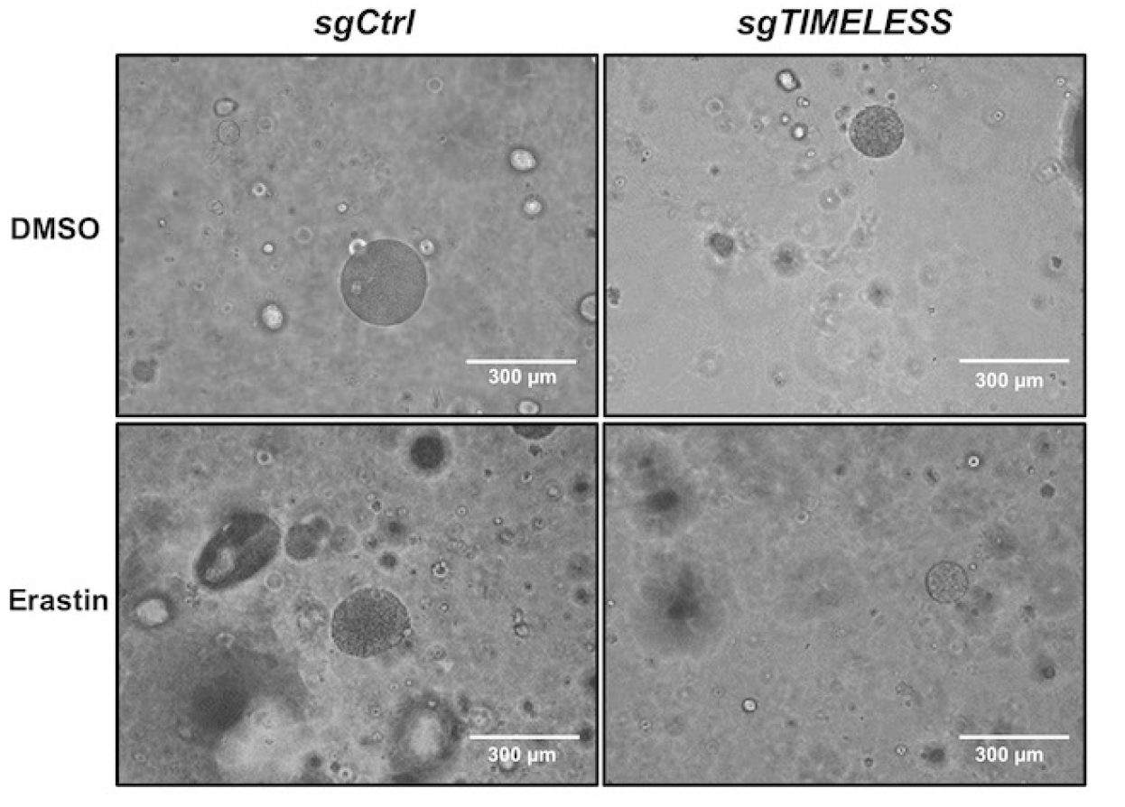
Figure 8. Representative bright‑field images of lung‑cancer organoids. TIMELESS knockout enhances Erastin‑mediated cytotoxicity in patient‑derived lung‑adenocarcinoma organoids. Scale bar = 300 μm.
2. Bioactive Materials, 2026 May 22. DOI:10.1016/j.bioactmat.2026.05.026. IF = 20.3
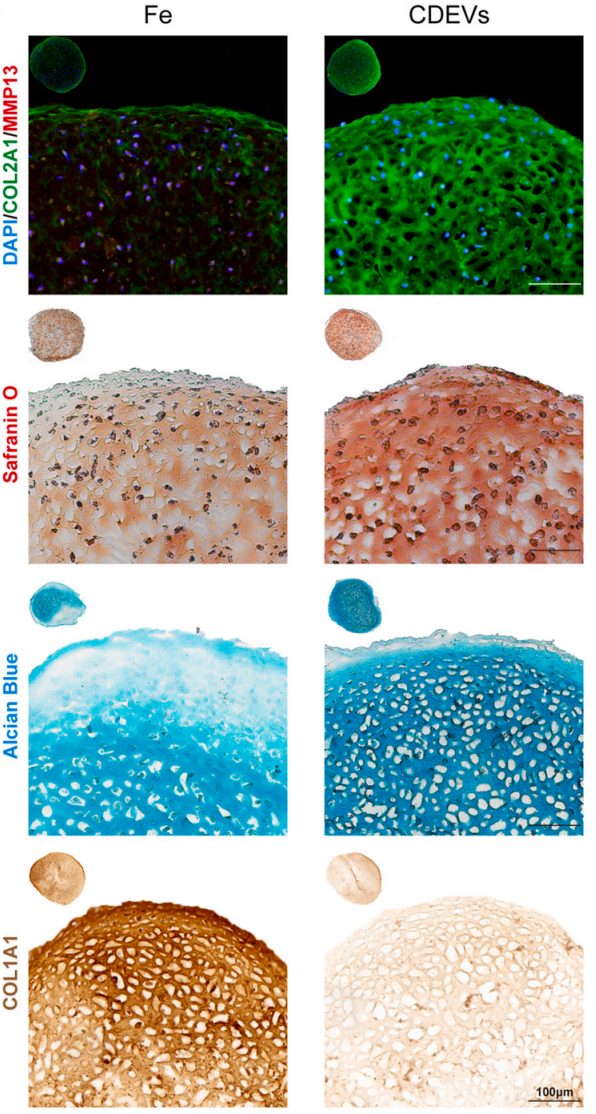
Study: Curcuma‑derived nanovesicle‑loaded ROS‑responsive hydrogels reprogram iron metabolism and improve cartilage regeneration post‑microfracture. Bone‑marrow‑derived mesenchymal‑stem‑cell (BMSC)‑based 3D cartilage microspheres were generated.
Reagent applied: abs9495 Low‑Growth‑Factor Phenol‑Red‑Free Matrix for BMSC‑derived 3D cartilage microsphere assembly.
3. Nature Communications, 2024 Feb 3. DOI:10.1038/s41467‑024‑45101‑9. IF = 16.6
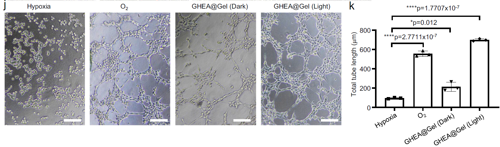
Study: Programmable microalgae‑integrated hydrogels promote diabetic chronic‑wound healing. The HEA@Gel formulation exhibits non‑cytotoxicity and generates oxygen to rescue cell viability under high‑glucose hypoxic conditions for skin‑cell and endothelial‑cell models.
Reagent applied: abs9493 High‑Concentration Phenol‑Red‑Free Matrix for HUVEC angiogenesis assays.
AntBio Key Product Lines


