UniOne® TR-FRET Human CDK2/CRBN PROTAC Binding Kit
UniOne® TR-FRET Human CDK2/CRBN PROTAC Binding Kit

Product Details
Product Details
Product Specification
| Host | Human |
| Stability & Storage | -80℃ |
Background
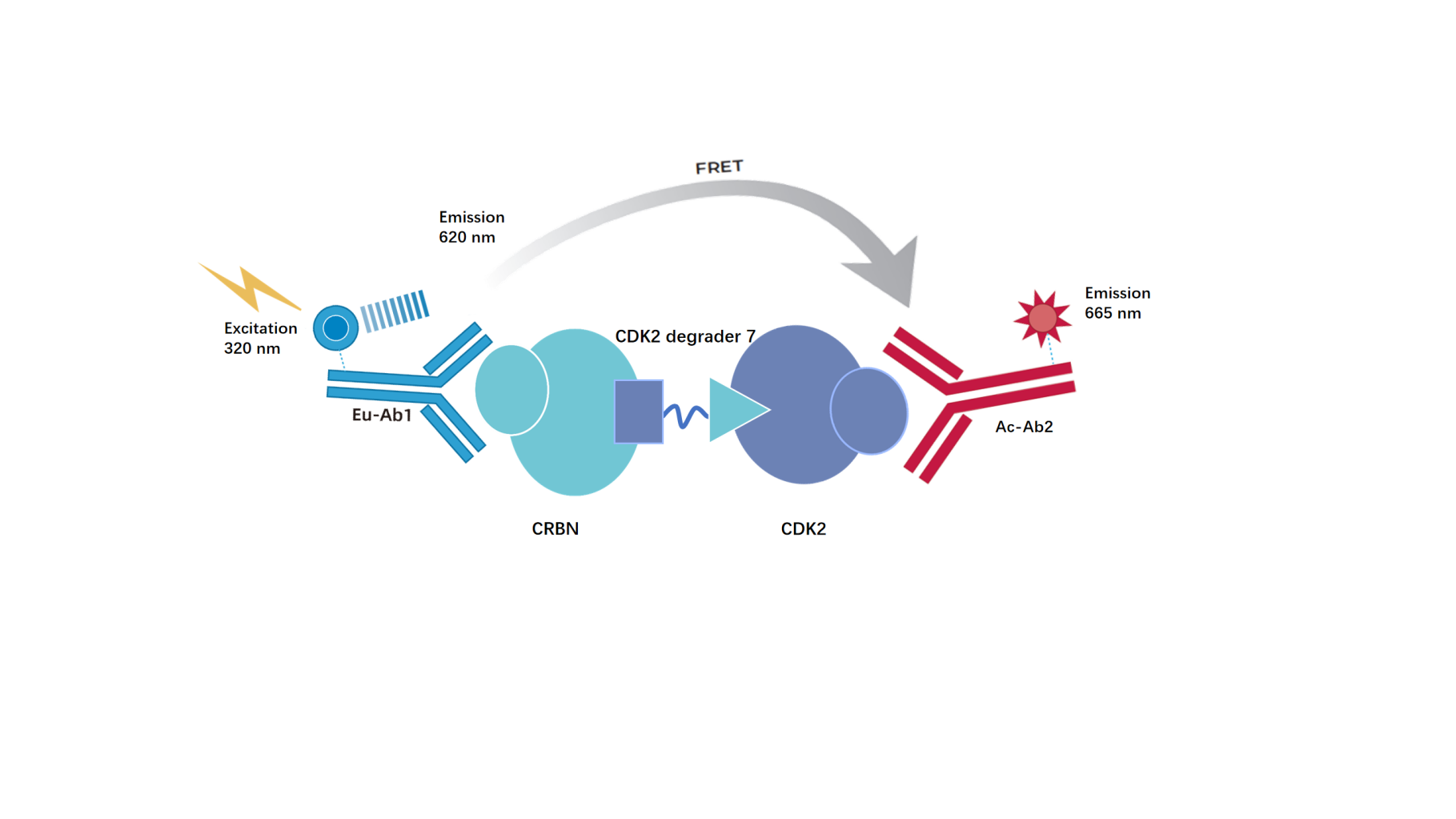
The kit employs homogeneous time-resolved fluorescence resonance energy transfer (TR‑FRET) technology for the quantification of molecular glue.CDK2 Degrader7and mediated by the compound to be detectedCRBN(DDB1/CRBN complex)withCDK2( CDK2/Cyclin E1 complex)Interaction. This method is simple and fast to implement.landThe assay is capable of mediating the DDB1/CRBN complex withCDK2High-throughput screening of small-molecule interactions.
AsThe following figureAs shown,CRBNandCDK2The interaction between them was detected using an Eu-labeled anti-Tag1 antibody (TR‑FRET donor) and an Ac‑labeled anti-Tag2 antibody (TR‑FRET acceptor). Due to the molecular glueCDK2 Degrader7Mediated CRBN andCDK2of the interaction, when the donor antibody approaches the receptor antibody, excitation of the donor antibody triggers fluorescence resonance energy transfer (FRET) to the receptor antibody, causing the receptor antibody to emit a signal at a wavelength of 665 nm. This specific signal is correlated withCDK2 Degrader7and CRBN,CDK2the extent of their interaction is directly proportional. This homogeneous assay is easy to perform and requires no washing.
Components
Product Componentsand Storage Conditions]
Components |
Concentration |
100T |
500T |
2500T |
10000T |
Storage Temperature |
Tag1-CRBNprotein |
100× |
5μL |
20μL |
100μL |
400μL |
-80℃ |
Tag2-CDK2protein |
100× |
5μL |
20μL |
100μL |
400μL |
-80℃ |
CDK2 Degrader 7 |
1mM |
5μL |
5μL |
25μL |
100μL |
-80℃ |
Anti-Tag1 Eu antibody |
50× |
5μL |
50μL |
250μL |
1mL |
-80℃ |
Anti-Tag2 Ac antibody |
100× |
5μL |
25μL |
125μL |
500μL |
-80℃ |
Detection buffer |
10× |
400μL |
2mL |
10mL |
40mL |
-80℃ |
Note: After the first thaw, aliquot immediately and store at the recommended temperature. Avoid storing diluted samples or subjecting them to repeated freeze-thaw cycles.
Protocol
1,Reagent Preparation
1.1Before use, allow all reagents to thaw at room temperature (equilibrate at room temperature for at least 30 minutes). The reaction volume in a 384‑well plate is 20 µL.muL (reagent volumes for the reaction system are shown in the table). Calculate the required reaction volume before preparation and prepare accordingly. The following formulation is for reference only, using 500 reactions as an example.
Table 1.Reagent Preparation
Reagent name |
Configuration |
μL per test well |
Detection buffer |
take2 mL 10timesDetection buffer1Join18 mL deionized water, diluteExplanationTo1×, mixedEvenly preparedUse. |
- |
CDK2 Degrader7 |
According to the reaction system, the compound is used withDilute the 1× detection buffer to the desired concentration.. |
2 |
Tag1-CRBN protein |
take20 μLTag1-CRBN proteinStock solution, 1× Detection buffer diluted to 2 mL,Mix well and set aside. |
4 |
Tag2-CDK2protein |
take20 μLTag2-CDK2proteinStock solution, diluted with 1× Detection buffer to 2 mL,mix well and set aside. |
4 |
Antibody Mix |
Take50μL Anti-Tag1 Eu antibodystock solution, add1× Detection buffer 2.450mL, mix well;take25μL Anti-Tag2 Ac antibodystock solution, add1× Detection buffer 2.475mL, mix well;the twoare mixed 1:1 to form the Antibody Mix. |
10 |
1.2sample to be testedgradient dilution
usingCDK2 Degrader7as an example, the diluent is 1× Detection buffer,to reduce the impact of matrix effect interference, it is recommended to use a solution with the same matrix as the sample being tested; andthe sample to be tested should be adjusted according to its actual concentration.
Table2.CDK2 Degrader7gradient dilution(adjusted based on actual conditions)
|
CDK2 Degrader7 final concentration (nM) |
CDK2 Degrader7Preparation concentration (nM) |
Preparation method |
|
① |
1000 |
10000 |
1μL1mMCDK2 Degrader7 +99μL 1× Detection buffer |
② |
333.3333 |
3333.333 |
20μL①+40μL 1× Detection buffer |
③ |
111.1111 |
1111.111 |
20μL②+40μL 1× Detection buffer |
④ |
37.0370 |
370.370 |
20μL③+40μL 1× Detection buffer |
⑤ |
12.3457 |
123.457 |
20μL④+40μL 1× Detection buffer |
⑥ |
4.1152 |
41.152 |
20μL⑤+40μL 1× Detection buffer |
⑦ |
1.3717 |
13.717 |
20μL⑥+40μL 1× Detection buffer |
⑧ |
0.4572 |
4.572 |
20μL⑦+40μL 1× Detection buffer |
⑨ |
0.1524 |
1.524 |
20μL⑧+40μL 1× Detection buffer |
Blank |
0 |
0 |
40μL of 1× Detection buffer |
2,Sample addition and controls
Positive drug standard curve |
Sample to be tested |
BlankControl well |
NC |
|
2μL Gradient dilutionCDK2 Degrader7 |
2μL Gradient-diluted sample to be tested |
2μL 1× Detection buffer |
10μL 1×Detection buffer Add10μL Antibody Mix |
4μLTag1-CRBN protein | |||
4μLTag2-CDK2protein | |||
10μL Antibody Mix | |||
Seal the plate wells with a sealing film.,Incubate at room temperature for 2 hours; | |||
2.1Sample to be tested: 4 μLTag1-CRBN proteinworking solution, 4 μLTag2-CDK2proteinworking solution, 2μLgradient dilutionsample to be tested,10μL of mixed Antibody Mix, added sequentially into the 384-well plate.
2.2 positive drug standard curve: 4μLTag1-CRBN proteinworking solution, 4μLTag2-CDK2 proteinworking solution,2μL gradient-dilutedCDK2 Degrader7, 10μL Antibody Mix.
2.3blankcontrolwell: 2μL of 1× Detection buffer insteadof the sample to be tested;
2.4NC: 10μL 1× Detection bufferadd10μL Antibody Mix.
After adding all samples, centrifuge, seal with a plate cover, and incubate at room temperature for 2 hours.
3,Detection
Detect on an ELISA reader compatible with TR-FRET..The excitation light is 320/340 nm, and the emission wavelengths are detected at 620 nm and 665 nm.Emissionwavelengths.
[Result Calculation]
1)Calculate the signal value(Ratio): Multiply the 665 nm fluorescence signal by the 620 nm fluorescence signal, then multiply by 10,000.
Ratio = (665/620)×10,000
2)Calculate based on the signal valueNet signal:
Net signal= (Std-NC)/NC×100
3)Calculate CV (%):
CV (%) = Standard Deviation / Mean Ratio × 100%
[[Data Example]
The following data cannot replace the data obtained in experiments; it is only for illustration, and results mayvary depending on the plate readerandequipment.
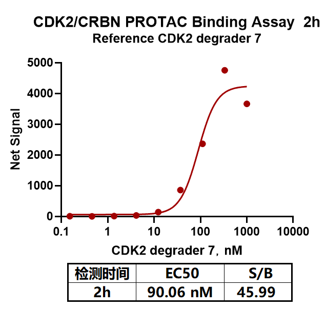
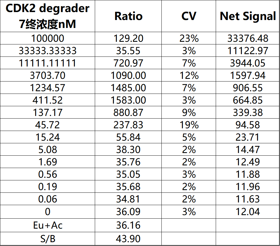
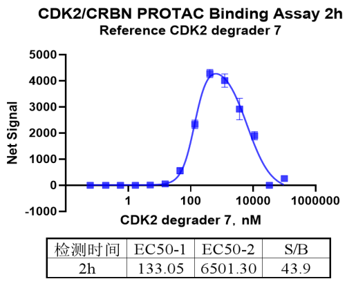
Note: RecommendedMicroplate (384-well plate, white, shallow wells)