Mouse Monoclonal Antibody Screening Set (Flag-tag Antigen)
Mouse Monoclonal Antibody Screening Set (Flag-tag Antigen)

Product Details
Product Details
Product Specification
| Stability & Storage | Store at 2-8°C away from light; product shelf life is 12 months. |
Background
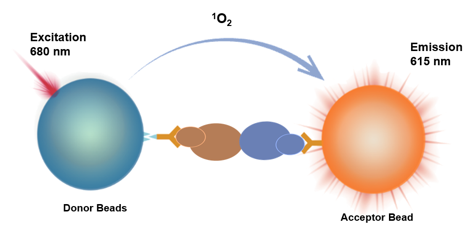
The Homogeneous Immuno Chemiluminescence Assay (HICA) is a homogeneous immunoassay method based on energy transfer between donor beads and acceptor beads at close proximity, resulting in luminescence.
Donor beads recognize Protein 1 (Tag1 label), while acceptor beads recognize Protein 2 (Tag2 label). When Protein 1 binds to Protein 2, the distance between the beads becomes less than 200nm. Upon excitation at 680nm, the donor beads generate singlet oxygen, which diffuses to the acceptor beads. The acceptor beads then undergo a redox reaction, emitting light at 615nm. The signal intensity is directly proportional to the strength of the protein interaction.
This product features a simple operation process, requires no washing, and offers high speed and sensitivity. It is capable of detecting weak interactions.
Components
Name |
Main Components |
3000T |
Reagent A |
Anti-Flag-tag antibody-crosslinked acceptor Beads |
10mL |
Reagent B |
Biotin-labeled anti-mouse IgG antibody |
10mL |
Reagent D |
Streptavidin Donor Beads |
10mL |
Protocol
[Reagents Required]
Name |
Catalog Number |
Universal Buffer 4 |
UA086115 |
[Test Procedure for Reference]
Test Procedure |
Test Procedure |
Step One: |
Add Flag-tag antigen to Reagent A at a concentration of 1.5 μg/mL. |
Step Two: |
Mix the above Reagent A, Reagent B, and Reagent D uniformly according to a volume ratio of 1:1:1.Protect from light/green light. |
Step Three: |
For each test, add 10 μL of the sample to be tested plus 10 μL of the mixed reagent.Protect from light/green light. |
Incubation. |
Incubate at 37°C with shaking for 60 minutes.Protect from light/green light. |
Reading. |
Instrument reading. |
[Sample and Reagent Preparation]
•Use DMEM+10%FBS to pre-dilute the mouse anti-IL1B antibody to 1000 ng/mL (6.67 nM) as C8, then perform gradient dilutions according to the following scheme:
Number |
Concentration (ng/mL) |
Dilution liquid volume (μL) |
Antibody volume (μL) |
C8 |
1000 |
/ |
/ |
C7 |
200 |
160 |
40 μL C8 |
C6 |
40 |
160 |
40 μL C7 |
C5 |
8 |
160 |
40 μL C6 |
C4 |
1.6 |
160 |
40 μL C5 |
C3 |
0.32 |
160 |
40 μL C4 |
C2 |
0.064 |
160 |
40 μL C3 |
C1 |
0 |
160 |
/ |
[Performance Verification]
•Reaction curve:
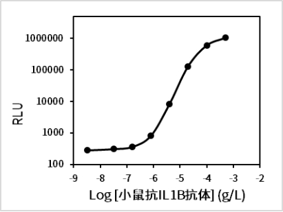
Highest signal: 1048745
Lowest signal: 280
EC50 = 2.106 ng/mL (0.014 nM)
•Blank limit:
Perform 20 repeated tests on C1, calculate the mean signal and SD, then use the reaction curve to determine the concentration value corresponding to mean signal + 2 × SD—this is the blank limit.
Blank limit = 0.738 ng/mL (4.922 pM)
•Repeatability:
High- and low-concentration samples were tested in duplicate ten times, and the concentration CV% was calculated.
Repeatability |
Low concentration |
High concentration |
CV% |
1.3% |
3.3% |
•Specificity: The following proteins were diluted to 1 μg/mL using DMEM+10%FBS, and the cross-interference rate was tested.
Tested substance |
Cross-reaction rate |
Human anti-IL1B antibody |
0.00% |
Guidelines
1. This experiment is light-sensitive. Please protect it from light during operation. It is recommended to perform preparation, sample addition, and incubation steps under green light (illuminance below 100 LUX).
2. This product is compatible with multi-functional microplate readers equipped with Alpha detection modules.
3. To ensure comparability of experimental data across different batches, please strictly control incubation temperature and time.
4. Avoid generating bubbles during sample addition.