

Mouse IgG2a Detection Kit [Wide Range] (HICA)
Mouse IgG2a Detection Kit [Wide Range] (HICA)

Product Details
Product Details
Product Specification
| Stability & Storage | Store at 2~8°C protected from light for 12 months; |
Background
This kit is used for the quantitative detection of mouse immunoglobulin G2a (mIgG2a) concentration in buffer solutions, cell culture supernatants, or serum samples.
Mouse immunoglobulin G2a consists of two heavy chains and two light chains. As a subclass with strong effector functions, mIgG2a can effectively activate the complement system and potently mediate antibody-dependent cell-mediated cytotoxicity (ADCC) and phagocytosis, playing a key role in processes such as antiviral immunity and tumor immune surveillance.
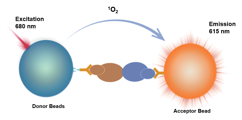
This kit employs a homogeneous luminescence competitive assay for the detection of mIgG2a concentration. Homogeneous luminescence is an immunoassay method based on energy transfer between donor and acceptor beads to produce luminescence.
It uses acceptor beads conjugated with anti-mIgG2a antibody (R1), biotin-labeled mIgG2a (R2), and donor beads conjugated with streptavidin (R3) to measure the concentration of mouse IgG2a.
When the test sample does not contain mIgG2a, R2 and R1 are mixed with the sample, and the biotin-labeled mIgG2a binds to the acceptor beads to form an immune complex, which then binds to the donor beads of R3 to form a luminescent complex. At this point, the distance between the two beads is less than 200 nm. Upon excitation, the donor beads produce singlet oxygen, which diffuses to the acceptor beads, and the acceptor beads generate corresponding emission light upon receiving the energy.
Conversely, when the test sample contains mIgG2a, the mIgG2a in the sample competitively binds to R1 with R2 to form an immune complex. After adding R3, the formation of luminescent complexes between donor and acceptor beads decreases. Upon laser excitation, the light signal produced by the acceptor beads will correspondingly decrease.
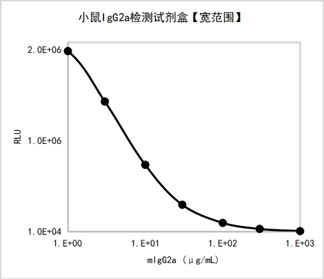
By collecting the light signal with the instrument's photosensitive element and using calibrators to fit a standard curve, the concentration of mIgG2a in the test sample can be calculated.
Components
Name |
Active Ingredient |
Content |
mIgG2aDetection ReagentR1 |
Receptor Microspheres Conjugated with Anti-Mouse IgG2a Antibody |
2mL/bottle × 1 |
mIgG2aDetection ReagentR2 |
Biotin-Labeled Mouse IgG2a |
2mL/bottle × 1 |
mIgG2aDetection ReagentR3 |
Donor Microspheres Conjugated with Streptavidin |
5mL/bottle × 1 |
mIgG2a Calibrator |
Mouse IgG2a |
100μg,Lyophilized Powder |
mIgG2a Calibrator Buffer |
BSA |
2mL/bottle × 1 |
Protocol
Materials and instruments required but not provided with this product:
•Luminometer/plate reader
•Multifunctional microplate reader with Alpha module
[Sample Requirements]
•Cell supernatant must be centrifuged at 1000 ×g for 10 minutes to remove particles and polymers.
•If the concentration of the sample to be tested exceeds the upper limit of detection, it is recommended to dilute before testing.
[Reference Testing Procedure]
Testing Procedure |
Testing Procedure (37℃) |
Testing Procedure (Room Temperature) |
Step One: |
Add 2 μL of calibrator/sample + 4 μL R2* + 4 μL R1 + 10 μL R3,Protect from light/green light |
Add 2 μL of calibrator/sample + 4 μL R2* + 4 μL R1 + 10 μL R3,Protect from light/green light |
Step Two: |
Incubate at 37℃ for 30 minutes,Protect from light/green light |
Incubate at room temperature for 60 minutes,Protect from light/green light |
Reading |
Instrument Reading |
Instrument reading |
*This kit is for competitive assay. Reagent R2 will react directly with R1, so they must not be pre-mixed before adding; also, pay attention to the order of addition and strictly follow the procedure!
[Calibrator Gradient Sample Preparation]
Reconstitute and prepare calibrator gradient samples using the same matrix as the samples to be tested. For example, if the test sample is cell culture supernatant, use a medium that has not been used to culture cells to reconstitute and prepare the calibrator gradient samples.
Dissolve the lyophilized calibrator in 100 μL of the matrix, then use the matrix todilute the mIgG2a calibrator. The recommended dilution scheme is shown in the following table:
Gradient |
Concentration (μg/mL) |
High concentration of mIgG2a |
Matrix (μL) |
C8 |
1000 |
/ |
/ |
C7 |
300 |
21 μL C8 |
49 |
C6 |
100 |
7 μL C8 |
63 |
C5 |
30 |
7μL C7 |
63 |
C4 |
10 |
7μL C6 |
63 |
C3 |
3 |
7μL C5 |
63 |
C2 |
1 |
7μL C4 |
63 |
C1 |
0 |
— |
63 |
Example of a complete standard curve:
[Performance Parameters]
•Blank Limit: Repeatedly test calibration standard C1 twenty times, calculate the mean signal and SD, then use the standard curve to determine the concentration value corresponding to mean signal minus 2×SD—this is the blank limit.
Testing Procedure |
Matrix |
Blank Limit (μg/mL) |
Testing Procedure 1 |
Calibration Standard Buffer |
0.23 |
Testing Procedure 2 |
Calibration Standard Buffer |
0.54 |
•Dynamic Range: 0–1000 μg/mL.
•Repeatability: Test high- and low-concentration samples ten times each and calculate the concentration CV.
Testing Procedure |
Matrix |
Repeatability |
|
Low Concentration |
High Concentration |
||
Test Procedure 1 |
Calibration Standard Buffer |
1.25% |
1.49% |
Test Procedure 2 |
Calibrator Buffer |
2.78% |
2.40% |
•Accuracy: Test accuracy samples and calculate the deviation from the target value.
Testing Procedure |
Matrix |
Deviation |
|
Low Concentration |
High Concentration |
||
Testing Procedure 1 |
Calibrator Buffer |
-0.65% |
2.03% |
Testing Procedure 2 |
Calibrator Buffer |
-0.29% |
-1.31% |
•Specificity: Dilute the following cross-reactants to 100 μg/mL using calibrator buffer, and test the cross-interference rate.
Testing Procedure |
Cross-reactant |
Cross-reaction rate |
Testing Procedure 1 |
Human IgG |
13.14% |
Rabbit IgG |
1.00% |
|
Pig IgG |
0.38% |
Guidelines
The detection reagent R3 is light-sensitive. Avoid exposure to light during use, and it is recommended to perform sample addition and incubation under green light (illuminance < 100 LUX).
It is recommended to recalibrate for each detection, with 2-3 replicate tests for each concentration point of the standard.
Four-parameter (weighted 1/Y²) or cubic spline fitting is recommended for calculation.
Pay attention to the required incubation temperature and duration.
Incubation at 37°C is recommended to be performed in the HiLA homogeneous luminescence analyzer.
Components from different reagent kit batches should not be mixed.
![Mouse IgG2a Detection Kit [Wide Range] (HICA)](http://www.antbioinc.com/cdn/shop/files/AntBioImage_5e714a01-b05e-4779-8dc8-eb3a02dc5db0.png?v=1785916972&width=1445)