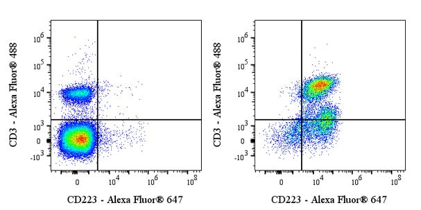
Flow cytometric analysis of mouse CD223 expression on mouse CD3 and CD28 antibody treated C57BL/6 mouse splenocytes. C57BL/6 mouse splenocytes untreated (left panel) or treated 72h with anti-mouse CD3, CD28 (right panel) were labeled with Mouse CD223 antibody at 1/500 dilution (1 μg). Goat Anti-Rat IgG Alexa Fluor® 647 was used as the secondary antibody. Then cells were stained with CD3 - Alexa Fluor® 488 antibody. Flow cytometry and data analysis were performed using Agilent NovoCyte Quanteon and FlowJo™ software.
Invivo anti-Mouse CD223 Recombinant mAb
Invivo anti-Mouse CD223 Recombinant mAb
Price:
Regular price
$170 USD
Regular price
Sale price
$170 USD
Unit price
per
For shipping services or bulk orders, you may request a quotation.
Secure checkout with

View full details
Product Details
Product Details
Product Specification
| Host | Rat |
| Antigen | CD223 |
| Synonyms | Lymphocyte activation gene 3 protein; LAG-3; Activation-induced cytidine deaminase-linked autoimmunity protein (Aida); Lag3 |
| Location | Secreted, Cell membrane |
| Accession | Q61790 |
| Clone Number | C9B7W |
| Antibody Type | Rat mAb |
| Isotype | Rat IgG1,k |
| Isotype Control | Invivo rat IgG1 Isotype control, anti-trinitrophenol |
| Application | FCM, Functional Assay, in vivo blocking, in vitro blocking |
| Reactivity | Ms |
| Positive Sample | Mouse CD3, CD28 antibody treated C57BL/6 mouse splenocytes |
| Purification | Protein G |
| Concentration | 5 mg/ml |
| Purity | >95% (Determined by SDS-PAGE) |
| Endotoxin | <1EU/mg |
| Conjugation | Unconjugated |
| Physical Appearance | Liquid |
| Storage Buffer | PBS pH7.4, containing no preservative |
| Stability & Storage | 2 to 8 °C for 2 weeks under sterile conditions; |
Dilution
| application | dilution | species |
| FCM | 1:500 | Ms |
Background
CD223, also known as LAG-3 (lymphocyte activation gene-3), is a type I transmembrane protein belonging to the immunoglobulin (Ig) superfamily. It is expressed on the surface of activated T cells, regulatory T cells (Tregs), natural killer (NK) cells, and some other immune cells. Structurally, it is similar to CD4 and has a higher binding affinity to MHC class II molecules than CD4. LAG-3 primarily functions as an immune checkpoint receptor, negatively regulating T-cell activation, proliferation, and cytokine production. In cancer and chronic infections, elevated LAG-3 expression is associated with T cell exhaustion, limiting anti-tumor and antiviral responses. Due to its significant role in immune regulation, LAG-3 has become a promising target for cancer immunotherapy and the treatment of autoimmune disorders.
Picture
Picture
FC